
Diffuse alveolar haemorrhage
Updates to Article Attributes
Diffuse alveolar haemorrhage (DAH) is a subset of diffuse pulmonary haemorrhage when bleeding is diffuse and directly into the alveolar spaces. It can occur in a vast number of clinical situations and at time can be life threatening.
Pathology
Blood tends to fill alveolar spaces as multiple sites.
Causes
It can occur with a number of causes which include:
-
pulmonary vasculitides (particularly small vessel vasculitides): a pulmonary capillaritis is considered the most common underlying lesion associated with diffuse alveolar haemorrhage
- ANCA associated pulmonary vasculitides
- non ANCA associated pulmonary vasculitides: Goodpasture syndrome
- certain connective tissue disorders
- systemic lupus erythematosus 2-3: considered commonest associated connective tissue disorder to result in diffuse alveolar haemorrhage
- mixed connective tissue disease 8
- coagulative disorders
- medications: excessive antiplatelet therapy 9
- inhaled toxins
- conditions causing back pressure
- haemorrhage complicating multifocal infection
- haemorrhage complicating diffuse alveolar damage
- haemorrhage complicating widespread pulmonary malignancy
- post bone marrow transplantation1
- pulmonary haemosiderosis
- antiphospholipid syndrome 6
Radiographic features
Plain film: chest radiograph
The clinical context is crucial in image interpretation. The exact pattern may differ dependant on the underlying cause. In general, the typical feature on plain film during an acute diffuse alveolar haemorrhage is a diffuse infiltrative opacification pattern 5. At times there may a slight predilection towards the mid zones 5 with some apical sparing 8.
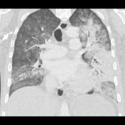
CT: HRCT Chest
The HRCT pattern can vary with time of onset of the haemorrhage and the clinical context is crucial in image interpretation.
-
acute phase
- can range from
lobularlobular or lobar areas of ground-glass opacities to predominant consolidation - ground-glass opacity is generated by subtotal alveolar filling with blood and is accompanied by apparent prominence of segmental and subsegmental bronchi, which has been referred to as the “dark bronchus sign"
- can range from
-
2–3 days
- intralobular lines and smooth interlobular septal thickening superimpose on areas of ground-glass opacity
- may sometimes give rise to a crazy-paving pattern 10
- these can often resolve
-
between chronic recurrent bleeding events
- ill-defined centrilobular nodules
- reflecting intra-alveolar accumulation of pulmonary macrophages
- usually
uniformuniform in size (1-3 mm) - diffusely distributed
- no zonal predominance
- ill-defined centrilobular nodules
- with severe repeated haemorrhage: may progress with features of interstitial fibrosis
Complications
Repeated episodes can lead to an organising pneumonia, collagen deposition in small airways and ultimately pulmonary fibrosis 7-8.
See also
-<a href="/articles/granulomatosis-with-polyangiitis">granulomatosis with polyangiitis </a>(formerly <a href="/articles/wegeners-granulomatosis">Wegener's granulomatosis</a>)</li>- +<a href="/articles/granulomatosis-with-polyangiitis">granulomatosis with polyangiitis </a>(formerly <a href="/articles/wegeners-granulomatosis">Wegener's granulomatosis</a>)</li>
-<li><a href="/articles/mitral-stenosis">mitral stenosis </a></li>- +<li><a href="/articles/mitral-stenosis">mitral stenosis </a></li>
-<li>post bone marrow transplantation <sup>1 </sup>- +<li>post bone marrow transplantation <sup>1 </sup>
-<li>can range from lobular or lobar areas of <a href="/articles/ground-glass-opacification">ground-glass opacities</a> to predominant consolidation </li>- +<li>can range from lobular or lobar areas of <a href="/articles/ground-glass-opacification">ground-glass opacities</a> to predominant consolidation </li>
-<li>usually uniform in size (1-3 mm) </li>- +<li>usually uniform in size (1-3 mm) </li>
-</ul><h4>Complications</h4><p>Repeated episodes can lead to an organising pneumonia, collagen deposition in small airways and ultimately pulmonary fibrosis <sup>7-8</sup>.</p><h4>See also</h4><ul><li><a href="/articles/pulmonary-haemorrhage">pulmonary haemorrhage </a></li></ul>- +</ul><h4>Complications</h4><p>Repeated episodes can lead to an organising pneumonia, collagen deposition in small airways and ultimately pulmonary fibrosis <sup>7-8</sup>.</p><h4>See also</h4><ul><li><a href="/articles/pulmonary-haemorrhage">pulmonary haemorrhage </a></li></ul>
Image 3 CT (lung window) ( create )


 Unable to process the form. Check for errors and try again.