
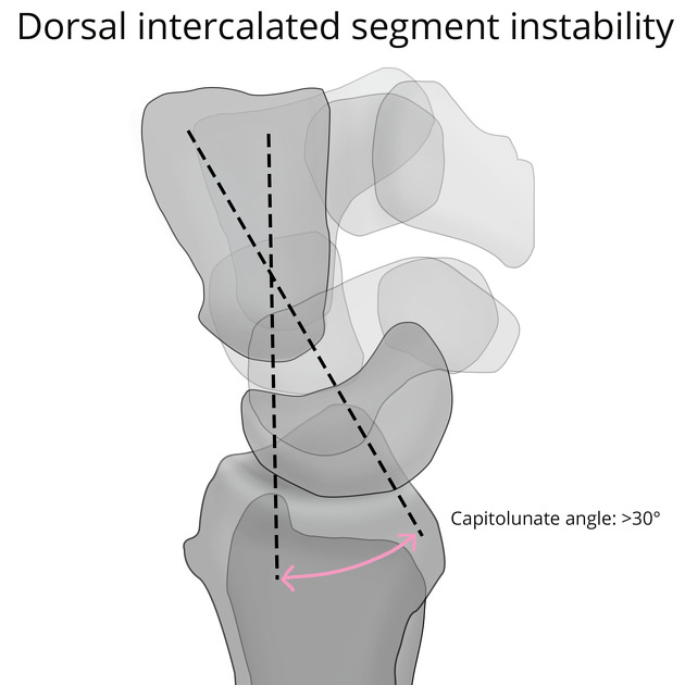
Dorsal intercalated segment instability (DISI) is a form of carpal instability featuring dorsal tilt of the lunate. It occurs mainly after the disruption of the scapholunate ligament and is more often encountered than volar intercalated segment instability (VISI).
On this page:
Clinical presentation
radial or dorsal wrist pain, maximal on radial deviation and wrist extension
weakness and/or instability
clicking wrist
positive Watson test: during ulnar to radial deviation, pressure applied to the volar aspect of the scaphoid elicits an audible and/or palpable clunk (due to dorsal subluxation of the scaphoid with respect to the radius)
Pathology
Etiology
-
wrist trauma, with or without a fracture
scaphoid fracture: bony DISI
distal radius fracture: compensatory DISI
radius malunion: adaptive DISI
scapholunate ligament dissociation: ligamentous DISI
Radiographic features
Fixed DISI deformity only occurs after combined injury of scapholunate ligament and other stabilizers of the scaphoid, namely radioscaphocapitate and scaphocapitate ligaments.
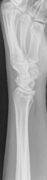
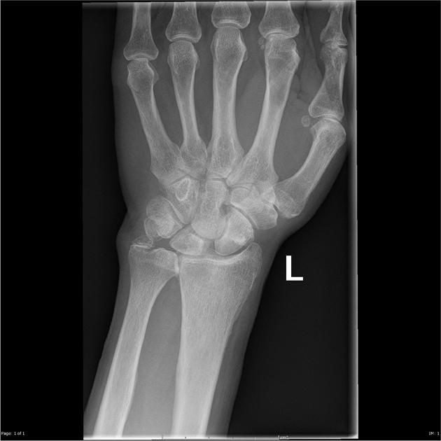
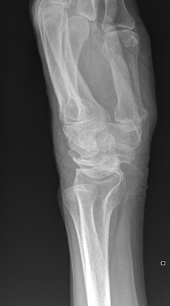
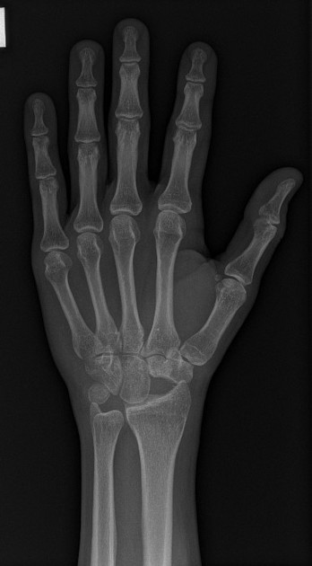
Plain radiograph
On an AP view, the normal trapezoidal configuration of the scaphoid may be lost and it may appear triangular.
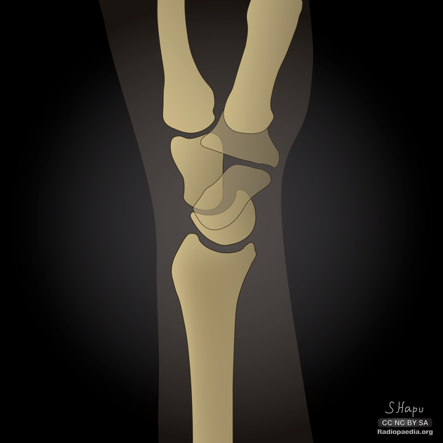
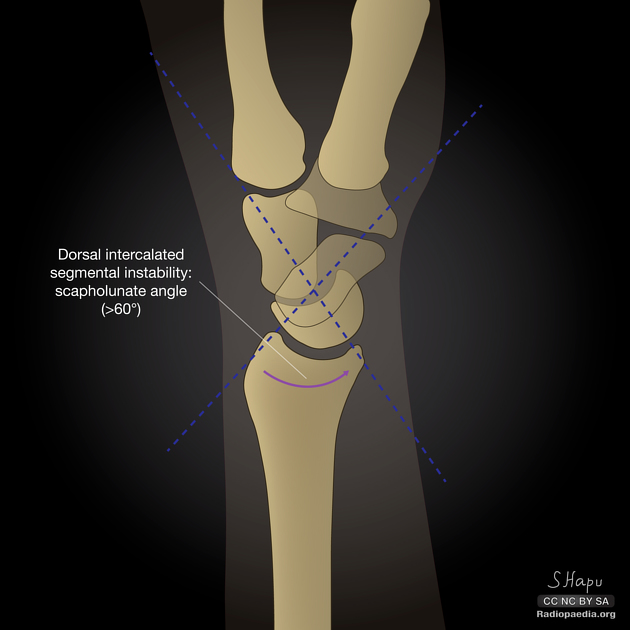
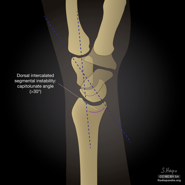
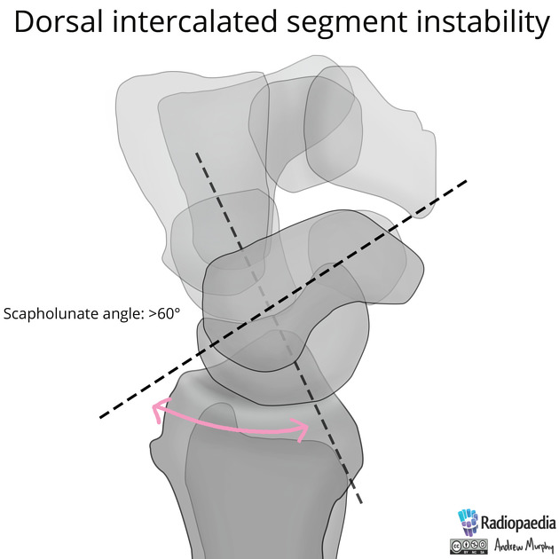
On the lateral radiograph with the wrist in a neutral position, DISI typically demonstrates dorsal tilt of the lunate with both of the following present:
scapholunate angle >60º: a sign of scapholunate ligament dissociation
capitolunate angle >30º: the capitate is displaced posteriorly compared to the distal radius
CT
On sagittal CT, the same findings as on lateral radiographs are seen.
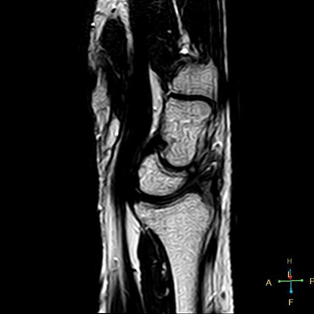
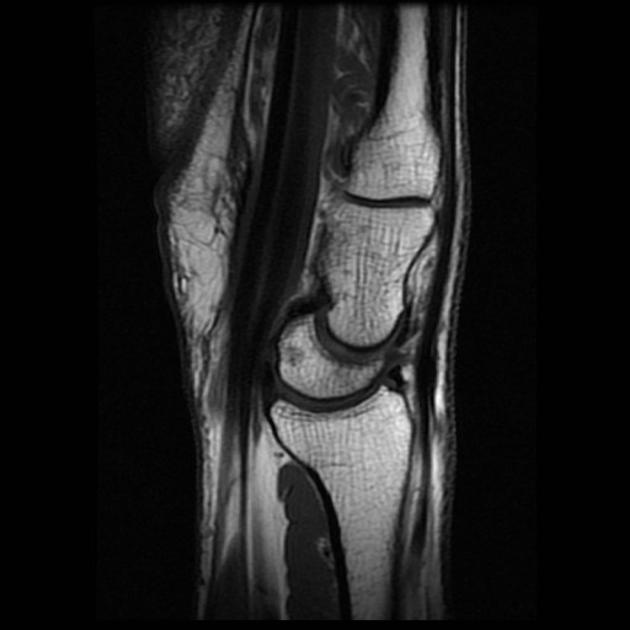
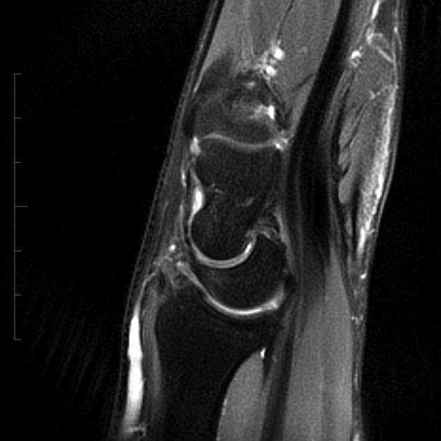
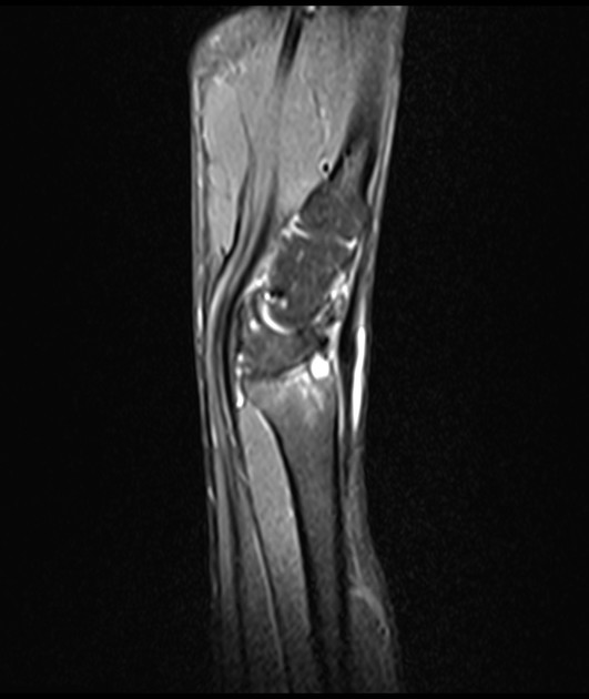
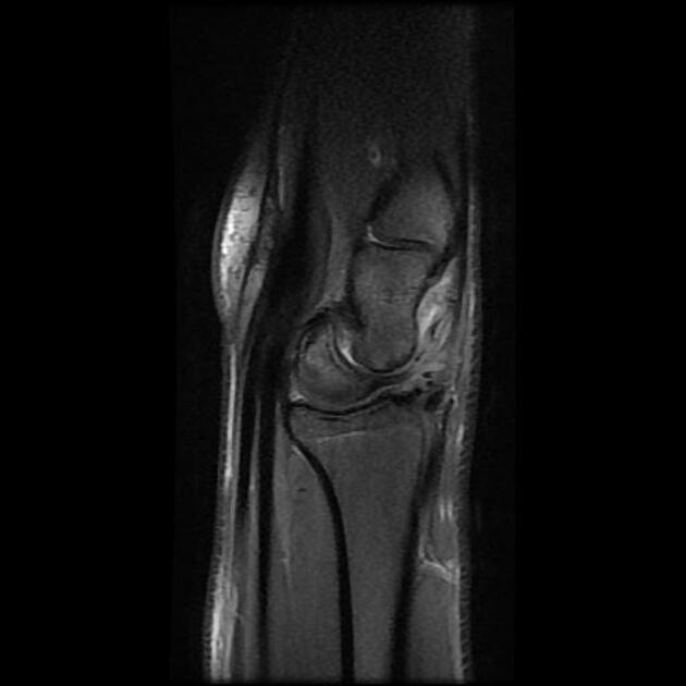
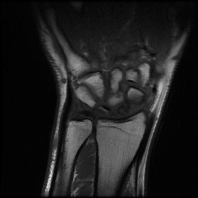
MRI
On sagittal MRI 9, the same findings as on plain radiograph are seen. However, bone contusion, scaphoid osteonecrosis, and scapholunate ligament injury are evaluated with more precision.


 Unable to process the form. Check for errors and try again.