
Epiphrenic diverticula are pulsion diverticula of the distal esophagus arising just above the lower esophageal sphincter, more frequently on the right posterolateral wall.
They are less frequent than traction mid esophageal diverticula but may have more clinical relevance.
On this page:
Clinical presentation
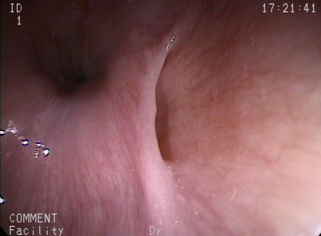
Epiphrenic diverticula may present with symptoms of dysphagia, and regurgitation. When large they can cause distal esophageal compression.
Pathology
These are considered to be pulsion type diverticula, thought to arise due to increased intraluminal pressure and hence the strong association with esophageal dysmotility.
The diverticulum forms as the submucosa and mucosa herniate focally through the muscularis propria. Since the diverticular wall lacks the muscular layer, it is classified as a "false" diverticulum.
Associations
They are associated with:
achalasia and other forms of neuromuscular dysfunction of the esophagus
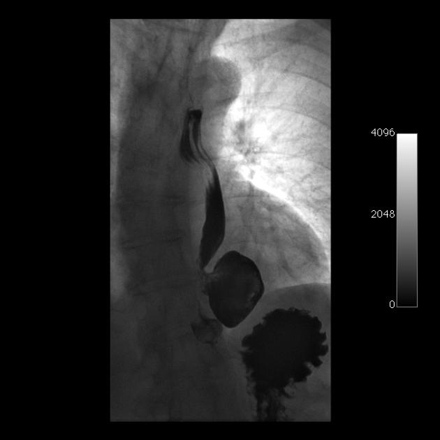
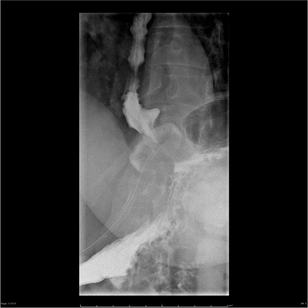
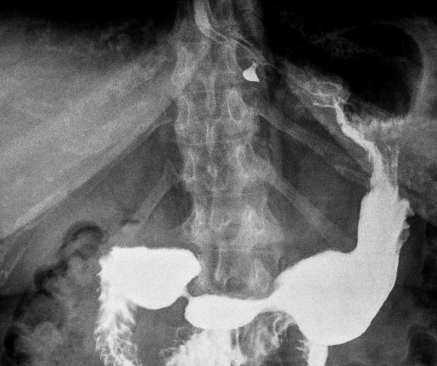
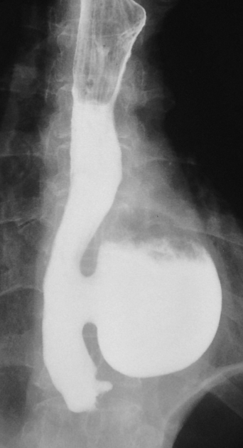
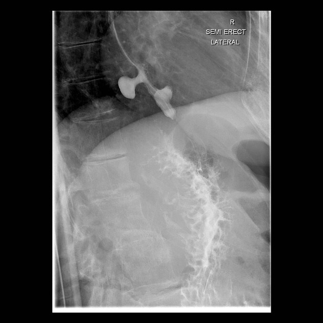
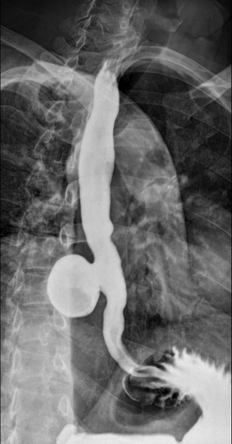
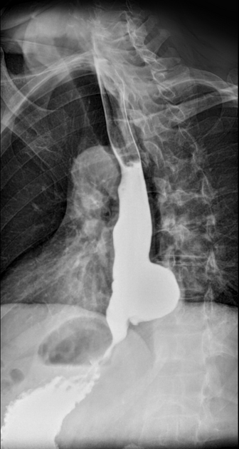
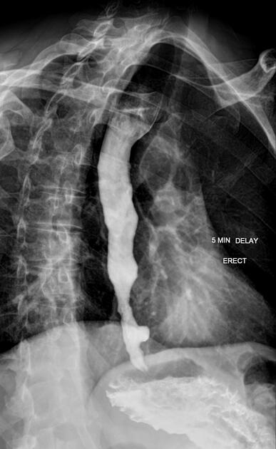
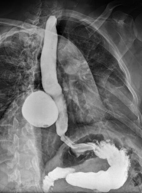
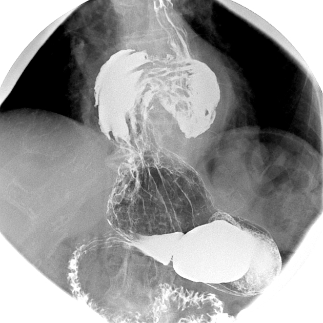
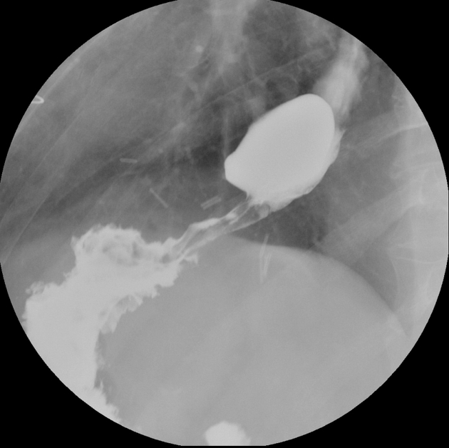
Radiographic features
Plain radiograph
On chest x-ray, they may appear as a retrocardiac soft tissue mass with or without an air-fluid level, mimicking a hiatus hernia.
Fluoroscopy
The best imaging method is a single contrast esophagogram, including prone RAO oblique views of the distal esophagus.
determine the relationship between the diverticulum and the gastro-esophageal junction
look for evidence of esophageal motility disorders and hiatus hernia
Treatment and prognosis
Conservative treatment can still be trialled for the treatment of an epiphrenic diverticulum 4. If incidentally found or in patients experiencing mild symptoms, conservative treatment and a period of observation can lead to complete resolution of symptoms. If symptoms persist, or in severely symptomatic patients with a large diverticulum, then surgical intervention is required 5. Surgical options include for epiphrenic diverticula include myomectomy or diverticulectomy.
Differential diagnosis
distal esophageal "ballooning" after an esophageal myotomy


 Unable to process the form. Check for errors and try again.