
Foreign body
Updates to Article Attributes
In medicine, foreignForeign bodies are objects lying partially or wholly within the body that originated in the external environment. Foreign body placement is voluntary or involuntary. Common voluntary acts will include cosmetic reasons, e.g. earrings (or other body piercings) and iatrogenic e.g. surgical clips, pacemaker; occasionally, however, the object has been inserted into natural cavities for sexual or nefarious purposes. Conversely, involuntary placement is usually as a result of an accident e.g. motor vehicle collisions, stepping on broken glass, gunshot wounds, or explosions 1-7 or perhaps inadvertently left behind after a procedure
Pathology
Aetiology
There are many ways in which a foreign body can be introduced into various parts of the human body.
Aspirated
Commonly, aspiratedAspirated foreign bodies will have a clear clinical correspondence: choking, coughing, neck pain or struggling to breathe. Risk factors for foreign body aspiration include intubation, neurological deficit, facial trauma, and dental instrumentation. The right main bronchus is the most common site of obstruction due to the anatomy of the bronchial tree favouring the right side (larger diameter, more vertical orientation). Often two orthogonal plain radiographs are the primary investigation of choice 12.
Iatrogenic
Many surgical procedures deliberately introduce foreign bodies either deliberately in the form of haemostatic clips (most common), stents, valve replacements, pacing boxes, catheters etc. or accidentally when a surgical instrument or surgical pack are inadvertently left in situ.
Ingested
Ingested foreign bodies range from children putting whatever they wantpaediatric patients placing objects in their mouths, to mental-health related issues of swallowing strange objects (pica)self-harm, to bones stuck in the pharynx or gastrointestinal tract. Coins account for 70% of paediatric ingested foreign bodies and will typically become ‘stuck’ at the level of the cricopharyngeus muscle 8,14. As a rule of thumb, coins visualised in the sagittal plane (acquired while entering through vocal cords) on anteroposterior radiographs are in the trachea, whereas coins in the oesophagus will have a coronal orientation on frontal chest radiographs.
An important alternative to consider when assessing coin-like objects are button batteries. These are very similar in appearance to coins, but typically have a slight step in profile with an inner ring when viewed en face. Button batteries can be potentially fatal when in contact with surrounding tissue as they can generate an electric current that will lead to the formation of sodium hydroxide resulting in severe, potentially fatal mucosal damage 6,13,14.
Sharp ingested foreign bodies can be potentially problematic when lodged in the oesophagus, patients will often require emergency endoscopy, more often than not if the sharp object is within reach of endoscopy it will be removed before it progresses further 12.Plastic bread clips are diagnostically challenging, the limited literature on this foreign body suggests they are invisible on both plain radiography and CT, and the rigid shape can cause bowel perforations or gastrointestinal haemorrhage 15,16.
Drug packing
Drug packing is a well-documented form of foreign body. Often drugs are placed within condoms, or wrapped in foil, latex or cellophane, then swallowed or inserted anally or vaginally. These should be considered hazardous to the patient until evacuated due to the possibility of the containment method rupturing.
Rectal
Most rectal foreign bodies are inserted via the anus, although occasionally the foreign body has been ingested and has passed through to lie in the rectum. Commonly, rectal foreign bodies are used for sexual purposes 1-3. Rectal foreign bodies are bewilderingly varied, ranging from manufactured sex toyswill vary case to improvised devices, vegetable, and a variety of improbable objects (e.g. light bulbs, and jars)case. The main problem with this is the often delayed presentation due to the reluctance of patients to present to emergency departments. Patients may suffer from extraperitoneal mucosal injuries or suffer from a more severe complication such as perforation 1-5.
Two plain radiographs are recommended to accurately demonstrate the size, shape and location of the rectal foreign body. This should be performed before a digital examination to prevent staff-related injuries from sharper foreign bodies.
Soft tissue
Most soft tissue foreign bodies are involuntary, resulting from an accident. The most commonly reported soft tissue foreign bodies are glass, metal and splinters from wood 5. It is imperative to locate foreign bodies before they become infected or worse damage close-by organs. Patients may have a delayed presentation with a foreign body granuloma.
Radiographic features
MetalThe investigation of foreign bodies relies heavily on radiology and every foreign body will have an optimal modality for investigation 17. Metal, glass and stone can be visualised very well using conventional plain film radiography, whereas more organic structures, such as wood, may require further imaging such as ultrasonography 5 5-7. Patients may have a delayed presentation with a foreign body granuloma.
Radiographic features
The investigation of foreign bodies relies heavily on radiology and every foreign body will have an optimal modality for investigation 17.
Plain radiographs
The radiological appearance in plain radiography of foreign bodies is dependent on three factors: the x-ray attenuation of the foreign body, the surrounding structures and any overlying structures that may veil the object.
The anatomical location will not only affect the radiopacity of the suspected foreign body, but the rate of magnification as the object is placed further or closer to the detector, lateral cervical radiographs can have a magnification rate of up to 21.6% 9-11.
Ultrasound
Most foreign bodies are hyperechoic.
CT
The density of the foreign material in the body can range markedly, so the use of multiple window settings is crucial in identifying the presence of the objects. Routinely soft tissue, lung, and bone windows should be assessed. Certain plastics may be occult on CT.
MRI
The signal intensity of the foreign material in the body can range markedly, but most objects cause artefact of some sort. Certain objects may be occult on MRI, such as those made of wood or plastic.
See also
-<p>In medicine, <strong>foreign bodies</strong> are objects lying partially or wholly within the body that originated in the external environment. Foreign body placement is voluntary or involuntary. Common voluntary acts will include cosmetic reasons, e.g. earrings (or other body piercings) and iatrogenic e.g. surgical clips, pacemaker; occasionally, however, the object has been inserted into natural cavities for sexual or nefarious purposes. Conversely, involuntary placement is usually as a result of an accident e.g. motor vehicle collisions, stepping on broken glass, gunshot wounds, or explosions <sup>1-7 </sup>or perhaps inadvertently left behind after a procedure.</p><h4>Pathology</h4><h5>Aetiology</h5><p>There are many ways in which a foreign body can be introduced into various parts of the human body. </p><h6>Aspirated</h6><p>Commonly, aspirated foreign bodies will have a clear clinical correspondence: choking, coughing, neck pain or struggling to breathe. Risk factors for foreign body aspiration include intubation, neurological deficit, facial trauma, and dental instrumentation. The right main bronchus is the most common site of obstruction due to the anatomy of the bronchial tree favouring the right side (larger diameter, more vertical orientation). Often two orthogonal plain radiographs are the primary investigation of choice <sup>12</sup>.</p><h6>Iatrogenic</h6><p>Many surgical procedures deliberately introduce foreign bodies either deliberately in the form of haemostatic clips (most common), stents, valve replacements, pacing boxes, catheters etc. or accidentally when a surgical instrument or surgical pack are inadvertently left in situ. </p><h6>Ingested</h6><p>Ingested foreign bodies range from children putting whatever they want in their mouths, mental-health related issues of swallowing strange objects (pica), to <a href="/articles/ingested-bones">bones stuck in the pharynx or gastrointestinal tract</a>. Coins account for 70% of paediatric ingested foreign bodies and will typically become ‘stuck’ at the level of the cricopharyngeus muscle <sup>8,14</sup>. As a rule of thumb, coins visualised in the sagittal plane (acquired while entering through vocal cords) on anteroposterior radiographs are in the <a href="/articles/trachea">trachea</a>, whereas coins in the<a href="/articles/oesophagus"> oesophagus</a> will have a coronal orientation on frontal chest radiographs.</p><p>An important alternative to consider when assessing coin-like objects are button batteries. These are very similar in appearance to coins, but typically have a slight step in profile with an inner ring when viewed en face. Button batteries can be potentially fatal when in contact with surrounding tissue as they can generate an electric current that will lead to the formation of sodium hydroxide resulting in severe, potentially fatal mucosal damage <sup>6,13,14</sup>.</p><p>Sharp ingested foreign bodies can be potentially problematic when lodged in the oesophagus, patients will often require emergency endoscopy, more often than not if the sharp object is within reach of endoscopy it will be removed before it progresses further <sup>12</sup>.<br><br>Plastic bread clips are diagnostically challenging, the limited literature on this foreign body suggests they are invisible on both plain radiography and CT, and the rigid shape can cause bowel perforations or gastrointestinal haemorrhage <sup>15,16</sup>.</p><h6>Drug packing</h6><p><a href="/articles/body-packing-1">Drug packing</a> is a well-documented form of foreign body. Often drugs are placed within condoms, or wrapped in foil, latex or cellophane, then swallowed or inserted anally or vaginally. These should be considered hazardous to the patient until evacuated due to the possibility of the containment method rupturing.</p><h6>Rectal</h6><p>Most <a href="/articles/rectal-foreign-bodies">rectal foreign bodies</a> are inserted via the anus, although occasionally the foreign body has been ingested and has passed through to lie in the rectum. Commonly, rectal foreign bodies are used for sexual purposes <sup>1-3</sup>. Rectal foreign bodies are bewilderingly varied, ranging from manufactured sex toys to improvised devices, vegetable, and a variety of improbable objects (e.g. light bulbs, and jars). The main problem with this is the often delayed presentation due to the reluctance of patients to present to emergency departments. Patients may suffer from extraperitoneal mucosal injuries or suffer from a more severe complication such as perforation <sup>1-5</sup>.</p><p>Two plain radiographs are recommended to accurately demonstrate the size, shape and location of the rectal foreign body. This should be performed before a digital examination to prevent staff-related injuries from sharper foreign bodies.</p><h6>Soft tissue</h6><p>Most soft tissue foreign bodies are involuntary, resulting from an accident. The most commonly reported soft tissue foreign bodies are <a href="/articles/glass-foreign-bodies">glass</a>, metal and splinters from wood <sup>5</sup>. It is imperative to locate foreign bodies before they become infected or worse damage close-by organs.</p><p>Metal, glass and stone can be visualised very well using conventional plain film radiography, whereas more organic structures, such as wood, may require further imaging such as ultrasonography<sup> 5-7</sup>. Patients may have a delayed presentation with a <a title="Foreign body granuloma" href="/articles/foreign-body-granuloma">foreign body granuloma</a>. </p><h4>Radiographic features</h4><p>The investigation of foreign bodies relies heavily on radiology and every foreign body will have an optimal modality for investigation <sup>17</sup>. </p><h5>Plain radiographs</h5><p>The radiological appearance in plain radiography of foreign bodies is dependent on three factors: the x-ray attenuation of the foreign body, the surrounding structures and any overlying structures that may veil the object. </p><p>The anatomical location will not only affect the radiopacity of the suspected foreign body, but the rate of magnification as the object is placed further or closer to the detector, lateral cervical radiographs can have a magnification rate of up to 21.6% <sup>9-11</sup>.</p><h5>Ultrasound</h5><p>Most foreign bodies are hyperechoic.</p><h5>CT</h5><p>The density of the foreign material in the body can range markedly, so the use of multiple window settings is crucial in identifying the presence of the objects. Routinely soft tissue, lung, and bone windows should be assessed. Certain plastics may be occult on CT.</p><h5>MRI</h5><p>The signal intensity of the foreign material in the body can range markedly, but most objects cause artefact of some sort. Certain objects may be occult on MRI, such as those made of wood or plastic.</p><h4>See also</h4><ul>- +<p><strong>Foreign bodies</strong> are objects lying partially or wholly within the body that originated in the external environment. Foreign body placement is voluntary or involuntary.</p><h4>Pathology</h4><h5>Aetiology</h5><p>There are many ways in which a foreign body can be introduced into various parts of the human body. </p><h6>Aspirated</h6><p>Aspirated foreign bodies will have a clear clinical correspondence: choking, coughing, neck pain or struggling to breathe. Risk factors for foreign body aspiration include intubation, neurological deficit, facial trauma, and dental instrumentation. The right main bronchus is the most common site of obstruction due to the anatomy of the bronchial tree favouring the right side (larger diameter, more vertical orientation). Often two orthogonal plain radiographs are the primary investigation of choice <sup>12</sup>.</p><h6>Iatrogenic</h6><p>Many surgical procedures deliberately introduce foreign bodies either deliberately in the form of haemostatic clips (most common), stents, valve replacements, pacing boxes, catheters etc. or accidentally when a surgical instrument or surgical pack are inadvertently left in situ. </p><h6>Ingested</h6><p>Ingested foreign bodies range from paediatric patients placing objects in their mouths, to mental-health related self-harm, to <a href="/articles/ingested-bones">bones stuck in the pharynx or gastrointestinal tract</a>. Coins account for 70% of paediatric ingested foreign bodies and will typically become ‘stuck’ at the level of the cricopharyngeus muscle <sup>8,14</sup>.</p><p>Sharp ingested foreign bodies can be potentially problematic when lodged in the oesophagus, patients will often require emergency endoscopy, more often than not if the sharp object is within reach of endoscopy it will be removed before it progresses further <sup>12</sup>.<br><br>Plastic bread clips are diagnostically challenging, the limited literature on this foreign body suggests they are invisible on both plain radiography and CT, and the rigid shape can cause bowel perforations or gastrointestinal haemorrhage <sup>15,16</sup>.</p><h6>Drug packing</h6><p><a href="/articles/body-packing-1">Drug packing</a> is a well-documented form of foreign body. Often drugs are placed within condoms, or wrapped in foil, latex or cellophane, then swallowed or inserted anally or vaginally. These should be considered hazardous to the patient until evacuated due to the possibility of the containment method rupturing.</p><h6>Rectal</h6><p>Most <a href="/articles/rectal-foreign-bodies">rectal foreign bodies</a> are inserted via the anus, although occasionally the foreign body has been ingested and has passed through to lie in the rectum. Commonly, rectal foreign bodies are used for sexual purposes <sup>1-3</sup>. Rectal foreign will vary case to case. The main problem with this is the often delayed presentation due to the reluctance of patients to present to emergency departments. Patients may suffer from extraperitoneal mucosal injuries or suffer from a more severe complication such as perforation <sup>1-5</sup>.</p><p>Two plain radiographs are recommended to accurately demonstrate the size, shape and location of the rectal foreign body. This should be performed before a digital examination to prevent staff-related injuries from sharper foreign bodies.</p><h6>Soft tissue</h6><p>Most soft tissue foreign bodies are involuntary, resulting from an accident. The most commonly reported soft tissue foreign bodies are <a href="/articles/glass-foreign-bodies">glass</a>, metal and splinters from wood <sup>5</sup>. It is imperative to locate foreign bodies before they become infected or worse damage close-by organs. Patients may have a delayed presentation with a <a href="/articles/foreign-body-granuloma">foreign body granuloma</a>. </p><h4>Radiographic features</h4><p>The investigation of foreign bodies relies heavily on radiology and every foreign body will have an optimal modality for investigation <sup>17</sup>. Metal, glass and stone can be visualised very well using conventional plain film radiography, whereas more organic structures, such as wood, may require further imaging such as ultrasonography<sup> 5-7</sup>. </p><h5>Plain radiographs</h5><p>The radiological appearance in plain radiography of foreign bodies is dependent on three factors: the x-ray attenuation of the foreign body, the surrounding structures and any overlying structures that may veil the object. </p><p>The anatomical location will not only affect the radiopacity of the suspected foreign body, but the rate of magnification as the object is placed further or closer to the detector, lateral cervical radiographs can have a magnification rate of up to 21.6% <sup>9-11</sup>.</p><h5>Ultrasound</h5><p>Most foreign bodies are hyperechoic.</p><h5>CT</h5><p>The density of the foreign material in the body can range markedly, so the use of multiple window settings is crucial in identifying the presence of the objects. Routinely soft tissue, lung, and bone windows should be assessed. Certain plastics may be occult on CT.</p><h5>MRI</h5><p>The signal intensity of the foreign material in the body can range markedly, but most objects cause artefact of some sort. Certain objects may be occult on MRI, such as those made of wood or plastic.</p><h4>See also</h4><ul>
-<li><a title="Foreign body granuloma" href="/articles/foreign-body-granuloma">foreign body granuloma</a></li>- +<li><a href="/articles/foreign-body-granuloma">foreign body granuloma</a></li>
Image ( destroy )
Image ( destroy )
Image ( destroy )
Image ( destroy )
Image ( destroy )
Image ( destroy )
Image ( destroy )
Image ( destroy )
Image ( destroy )
Image ( destroy )
Image ( destroy )
Image ( destroy )
Image ( destroy )
Image ( destroy )
Image ( destroy )
Image ( destroy )
Image ( destroy )
Image ( destroy )
Image ( destroy )
Image ( destroy )
Image ( destroy )
Image ( destroy )
Image ( destroy )
Image ( destroy )
Image ( destroy )
Image ( destroy )
Image ( destroy )
Image ( destroy )
Image ( destroy )
Image ( destroy )
Image ( destroy )
Image ( destroy )
Image ( destroy )
Image ( destroy )
Image ( destroy )
Image ( destroy )
Image ( destroy )
Image ( destroy )
Image ( destroy )
Image ( destroy )
Image ( update )
Image 2 X-ray ( update )

Image 3 CT (bone window) ( update )

Image 4 CT (non-contrast) ( update )

Image 5 X-ray (Frontal) ( update )

Image 6 CT (non-contrast) ( update )

Image 7 CT (bone window) ( update )

Image 8 X-ray ( update )

Image 9 X-ray (Lateral) ( update )

Image 10 X-ray (Lateral) ( update )

Image 11 CT (C+ CTPA) ( update )

Image 12 X-ray (Frontal) ( update )
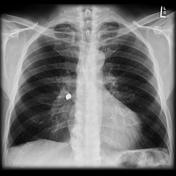
Image 13 CT (lung window) ( update )

Image 14 Mammography (MLO) ( update )
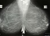
Image 15 Annotated image (Lateral) ( update )
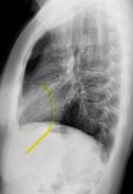
Image 16 X-ray (Frontal) ( update )

Image 17 X-ray (Frontal) ( update )
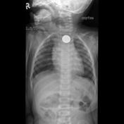
Image 18 X-ray (Lateral) ( update )

Image 19 X-ray (Frontal) ( update )

Image 20 CT (liver window) ( update )

Image 21 X-ray (AP supine) ( update )

Image 22 Annotated image (Axial) ( update )
Image 23 CT (3D) ( update )

Image 24 X-ray ( update )

Image 25 X-ray (Frontal) ( update )

Image 26 X-ray (Lateral) ( update )

Image 27 X-ray ( update )

Image 28 X-ray (Frontal) ( update )

Image 29 X-ray (Frontal) ( update )

Image 30 X-ray (Frontal) ( update )

Image 31 X-ray (Frontal) ( update )

Image 32 X-ray (Lateral) ( update )


 Unable to process the form. Check for errors and try again.