
Gliomatosis cerebri is an uncommon growth pattern of diffuse gliomas that involves at least three lobes by definition, has frequent bilateral growth and may extend to infratentorial structures 8. There often is an important discordance between clinical and radiological findings, as it may be clinically silent while it appears as a very extensive process radiologically.
Importantly, whereas gliomatosis was previously considered a distinct entity since the 2016 update to the WHO classification of CNS tumors it is now merely thought of as a growth pattern 8.
On this page:
Epidemiology
peak incidence is 20-40 years of age
M:F of 1.5:1 in one sequential series of 54 patients 7
Pathology
Gliomatosis cerebri growth pattern can be seen in all diffuse gliomas of any grade 6,7.
Classification
Gliomatosis cerebri can be divided into two types 7:
-
type 1: no discrete mass
usually IDH wild-type 8
-
type 2: discrete mass with further diffuse CNS involvement
IDH mutation is more common in this subtype 7
Radiographic features
CT
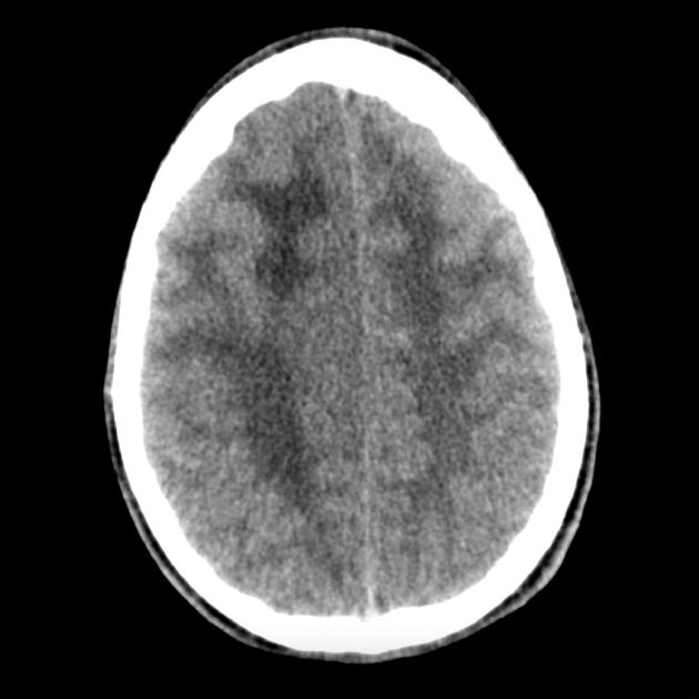
CT can be normal as lesions are often isodense to normal brain parenchyma. There is a relative lack of mass effect and distortion compared to the extensiveness of involvement. There may be an ill-defined asymmetry or subtle hypoattenuation of the involved brain parenchyma.
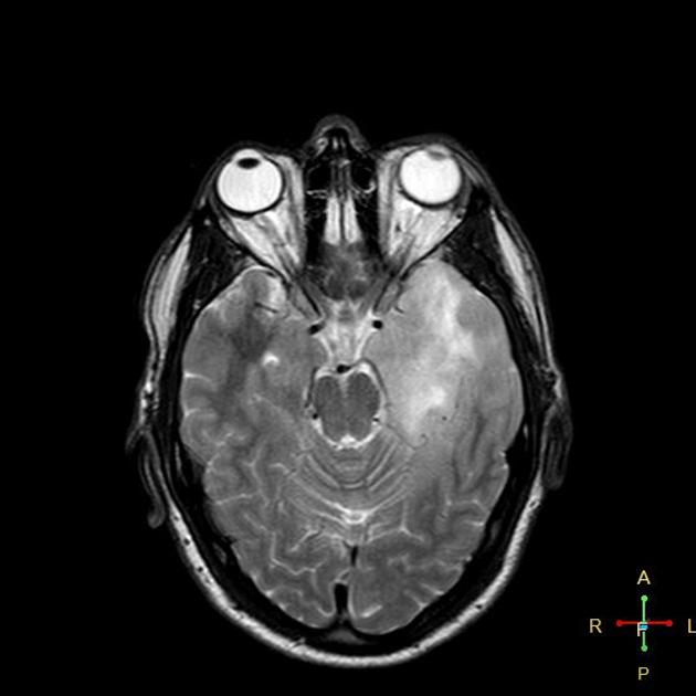
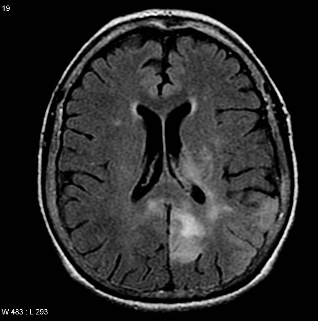
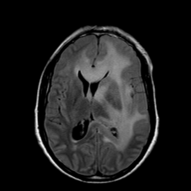
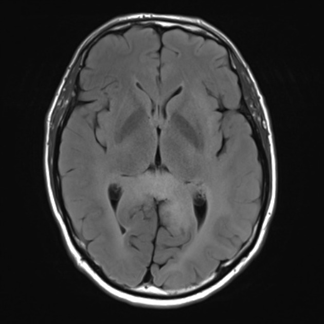
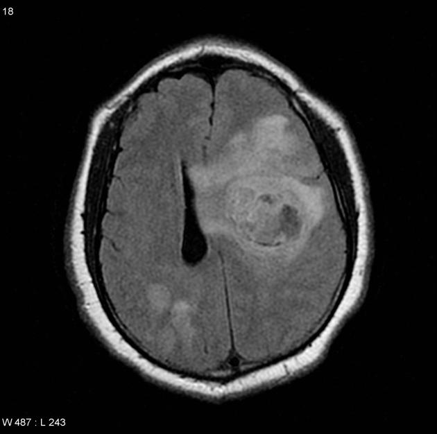
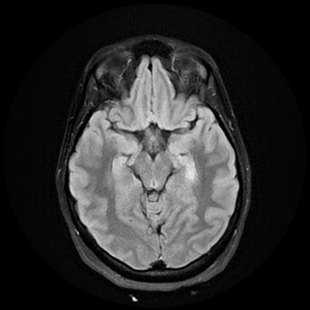
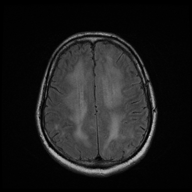
MRI
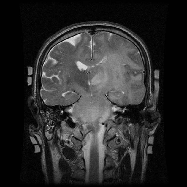
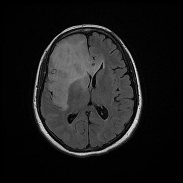
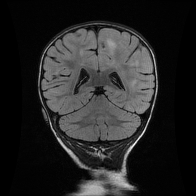
Mass effect and enhancement are often minimal despite large portions of the brain being involved. There is a loss of grey-white matter differentiation and diffuse gyral thickening.
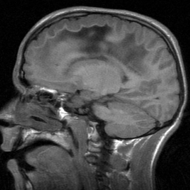
Diffuse T1 and T2 prolongation throughout both white and grey matter:
T1: iso to hypointense to grey matter 1
T2: hyperintense to grey matter 1
T1 C+ (Gd): typically no or minimal enhancement
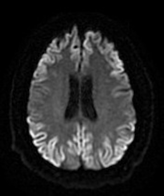
DWI: usually no restriction
-
MR spectroscopy
elevated Cho:Cr and Cho:NAA ratios 2
marked elevation of myo-inositol (mI)
-
perfusion MR
low/normal rCBV: correlates with no vascular hyperplasia
PET
FDG-PET shows marked hypometabolism
Treatment and prognosis
The condition carries a poor prognosis with an average survival of ~50% at 1 year and 25% at 3 years. Transformation into glioblastoma may occur a few years later.
Surgery is not a viable option due to the diffuse nature of this tumor but can be used to treat complications such as hydrocephalus or mass effect.
Radiation therapy has been shown to increase survival 7, however, the large field usually required in these lesions increases the risk of severe toxicity 5. Chemotherapy is a treatment option, although there is a lack of evidence in its use (both positive and negative) 7.
Differential diagnosis
General imaging differential considerations depend very much on the feature that is most prominent.
When widespread cortical involvement is prominent then it is worth considering:
-
less marked swelling
spontenous rapid resolution of imaging changes
-
subacute ischemia, global hypoxic-ischemic injury or hyperperfusion syndrome
presentation is usually quite different
-
the cortex is usually not swollen and subjacent white matter is normal
-
when viral the clinical presentation is usually far more acute
paraneoplastic/autoimmune encephalitis can be harder to distinguish


 Unable to process the form. Check for errors and try again.