
Hepatomegaly refers to an increase in size or enlargement of the liver.
On this page:
Pathology
Etiology
Hepatomegaly can result from a vast range of pathology including, but not limited to, the following:
-
malignancy/cellular infiltrate
-
acquired hepatic conditions
-
acquired non-hepatic conditions
-
congenital anomalies
-
biochemical
-
anatomical
-
-
syndromes
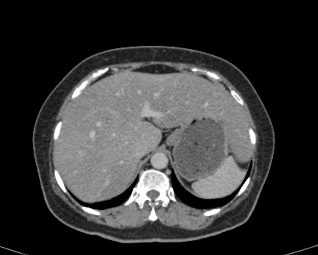
Radiographic features
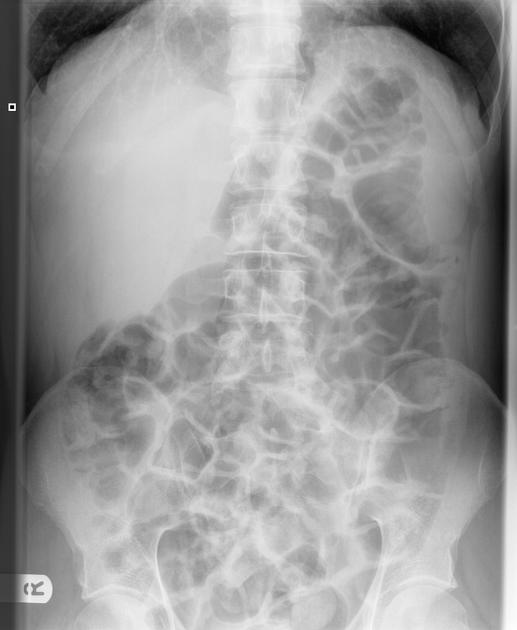
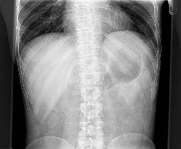
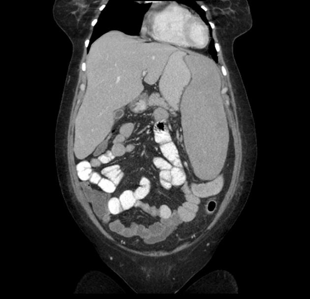
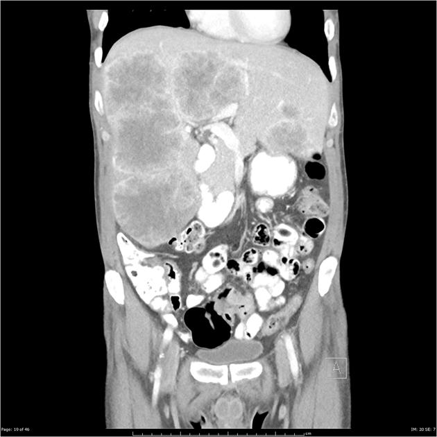
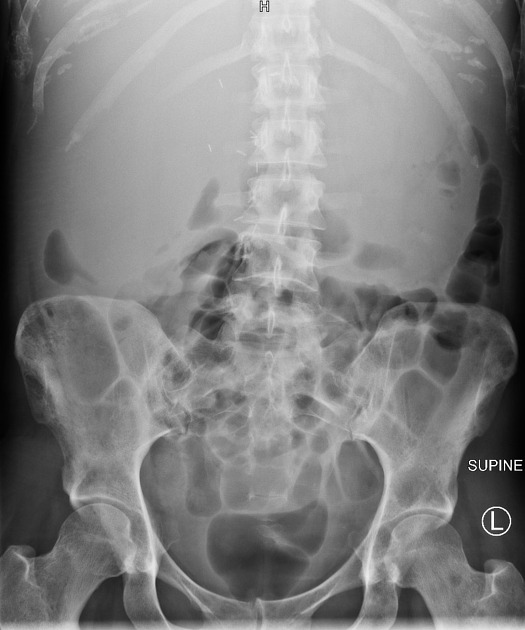
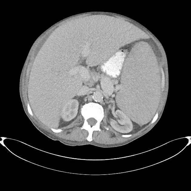
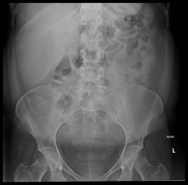
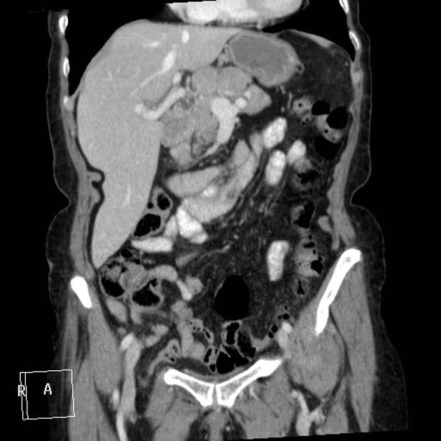
Assessment of liver size is commonly made on ultrasound or CT, although gross hepatomegaly may be apparent on abdominal radiograph.
For the adult liver:
-
midclavicular line averages 10-12.5 cm in craniocaudal length 2
a liver that is longer than 15.5-16 cm in the midclavicular line (MCL) is considered enlarged
average transverse length is 20-23 cm at the level of the upper pole of the right kidney 2
In practice, however, assessment is often subjective.
Features that support hepatomegaly include 1:
extension of the right lobe inferior to the lower pole of the right kidney
rounding of the hepatic inferior border
Liver volume can be assessed on cross-sectional imaging either using volumetry or by calculating an estimated liver volume from caliper measurements. The following formula was proposed for this purpose 4:
Volume = maximum cranio-caudal dimension x maximum latero-lateral dimension x maximum antero-posterior dimension x 0.31
The range of normal liver volume is however dependent on patient population and demographics. Furthermore, liver volume has been shown to demonstrate a diurnal rhythm due to hydration, nutrition, and physical activity, reaching its minimum value between 12:00-14:00 hours 5,6.


 Unable to process the form. Check for errors and try again.