
Hereditary multiple exostoses
Updates to Article Attributes
Hereditary multiple exostoses (HME), also known as diaphyseal aclasis or osteochondromatosis is an autosomal dominant condition, characterised by the development of multiple osteochondromas.
Epidemiology
Hereditary multiple exostoses demonstrate an autosomal dominant inheritance pattern, with incomplete penetrance in females. The number of exostoses, the degree, and type of angular deformity, and even the rate of malignant transformation varies significantly, even within families.
Clinical presentation
Most patients are diagnosed by the age of 5 years, and virtually all are diagnosed by the age of 12 years. Patients may be asymptomatic with a few small lesions or may be significantly deformed by multiple large osteochondromas.
Pathology
Location
It can involve any bony in the body except for the calvarium 5.
Common sites of involvement include the distal femur, proximal tibia, wrist and hands, humerus, ankle, pelvis and ribs.
Genetics
It is though to result from EXT1, EXT2 or EXT 3 gene mutationson chromosomes 8q24 (EXT1), 11p11-13 (EXT2) and 19p (EXT3) 4-5.
Radiographic features
Except that they are multiple, imaging features are identical to solitary osteochondromas. The skeletal distribution of lesions can significantly vary, with some authors noting that the typical distribution is bilateral and symmetric, whereas others report a strong unilateral predominance.
Often associated with a broadened shaft at the end of long bones, hence the term diaphyseal aclasis.
Complications
Are also similar to those of solitary osteochondroma and include:
- vascular impingement
- neural impingement
- fracture
- bursitis
- deformity and ankylosis
- malignant transformation
Malignant transformation is more common than in sporadic cases, with transformation rates reported as high as 25% (lower rates of 3-5% have also been published) 3. The mnemonic GLAD PAST 1 lists the associations with sarcomatous transformation.
Refer to the generic osteochondroma article for more information.
See also
-<p><strong>Hereditary multiple exostoses</strong>, also known as <strong>diaphyseal aclasis</strong> or <strong>osteochondromatosis</strong> is an <a href="/articles/autosomal-dominant">autosomal dominant</a> condition, characterised by the development of multiple <a href="/articles/osteochondroma">osteochondromas</a>.</p><h4>Epidemiology</h4><p>Hereditary multiple exostoses demonstrate an autosomal dominant inheritance pattern, with incomplete penetrance in females. The number of exostoses, the degree, and type of angular deformity, and even the rate of malignant transformation varies significantly, even within families.</p><h4>Clinical presentation</h4><p>Most patients are diagnosed by the age of 5 years, and virtually all are diagnosed by the age of 12 years. Patients may be asymptomatic with a few small lesions or may be significantly deformed by multiple large osteochondromas. </p><h4>Radiographic features</h4><p>Except that they are multiple, imaging features are identical to solitary <a href="/articles/osteochondroma">osteochondromas</a>. The skeletal distribution of lesions can significantly vary, with some authors noting that the typical distribution is bilateral and symmetric, whereas others report a strong unilateral predominance.</p><p>Often associated with a broadened shaft at the end of long bones, hence the term diaphyseal aclasis. </p><h4>Complications</h4><p>Are also similar to those of solitary <a href="/articles/osteochondroma">osteochondroma </a>and include:</p><ul>- +<p><strong>Hereditary multiple exostoses (HME)</strong>, also known as <strong>diaphyseal aclasis</strong> or <strong>osteochondromatosis</strong> is an <a href="/articles/autosomal-dominant">autosomal dominant</a> condition, characterised by the development of multiple <a href="/articles/osteochondroma">osteochondromas</a>.</p><h4>Epidemiology</h4><p>Hereditary multiple exostoses demonstrate an autosomal dominant inheritance pattern, with incomplete penetrance in females. The number of exostoses, the degree, and type of angular deformity, and even the rate of malignant transformation varies significantly, even within families.</p><h4>Clinical presentation</h4><p>Most patients are diagnosed by the age of 5 years, and virtually all are diagnosed by the age of 12 years. Patients may be asymptomatic with a few small lesions or may be significantly deformed by multiple large osteochondromas. </p><h4>Pathology</h4><h5>Location</h5><p>It can involve any bony in the body except for the calvarium <sup>5</sup>.</p><p>Common sites of involvement include the distal femur, proximal tibia, wrist and hands, humerus, ankle, pelvis and ribs.</p><h5>Genetics</h5><p>It is though to result from EXT1, EXT2 or EXT 3 gene mutations<sup> </sup>on chromosomes 8q24 (<em>EXT1</em>), 11p11-13 (<em>EXT2</em>) and 19p (<em>EXT3</em>) <sup>4-5</sup>.</p><h4>Radiographic features</h4><p>Except that they are multiple, imaging features are identical to solitary <a href="/articles/osteochondroma">osteochondromas</a>. The skeletal distribution of lesions can significantly vary, with some authors noting that the typical distribution is bilateral and symmetric, whereas others report a strong unilateral predominance.</p><p>Often associated with a broadened shaft at the end of long bones, hence the term diaphyseal aclasis. </p><h4>Complications</h4><p>Are also similar to those of solitary <a href="/articles/osteochondroma">osteochondroma </a>and include:</p><ul>
References changed:
- 4. Ellatif M, Sharif B, Lindsay D, Pollock R, Saifuddin A. An Update on the Imaging of Diaphyseal Aclasis. Skeletal Radiol. 2021;50(10):1941-62. <a href="https://doi.org/10.1007/s00256-021-03770-3">doi:10.1007/s00256-021-03770-3</a> - <a href="https://www.ncbi.nlm.nih.gov/pubmed/33791832">Pubmed</a>
- 5. Kok H, Fitzgerald L, Campbell N et al. Multimodality Imaging Features of Hereditary Multiple Exostoses. Br J Radiol. 2013;86(1030):20130398. <a href="https://doi.org/10.1259/bjr.20130398">doi:10.1259/bjr.20130398</a> - <a href="https://www.ncbi.nlm.nih.gov/pubmed/24004486">Pubmed</a>
Image ( update )
Image 1 Diagram ( update )

Image 2 X-ray (Frontal) ( update )

Image 4 X-ray (Frontal) ( update )
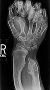
Image 5 X-ray ( update )
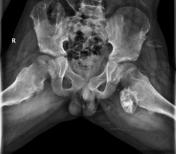
Image 6 X-ray (Frontal) ( update )
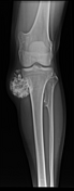
Image 7 X-ray (Frontal) ( update )

Image 8 X-ray ( update )
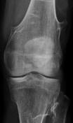
Image 9 X-ray ( update )
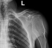
Image 10 X-ray ( update )

Image 11 X-ray (Frontal) ( update )

Image 12 X-ray (Frontal) ( update )

Image 13 X-ray (Frontal) ( update )

Image 14 Annotated image (AP annotated) ( update )
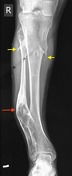
Image 16 X-ray (Frontal) ( update )

Image 17 X-ray (Frontal) ( update )

Image 18 CT (3D VRT) ( update )

Image 20 X-ray (Frontal) ( update )

Image 21 X-ray (Frontal) ( update )

Image 22 X-ray (Frontal) ( update )

Image 23 X-ray (Frontal) ( update )

Image 24 X-ray (Knees frontal ) ( update )
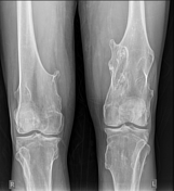
Image 25 X-ray (CHEST X-RAY-FRONTAL) ( update )

Image 26 CT (C+ portal venous phase) ( update )

Image 27 X-ray (Frontal) ( update )

Image 28 X-ray (Frontal) ( update )


 Unable to process the form. Check for errors and try again.