
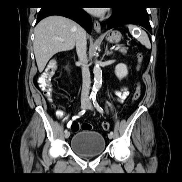
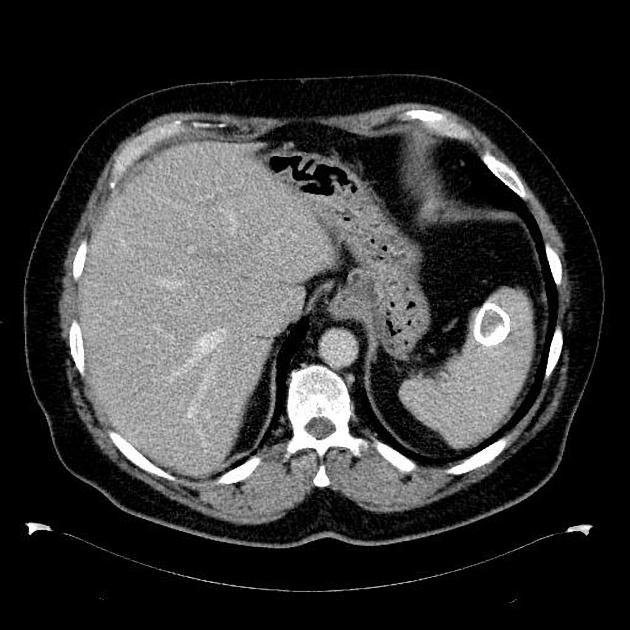
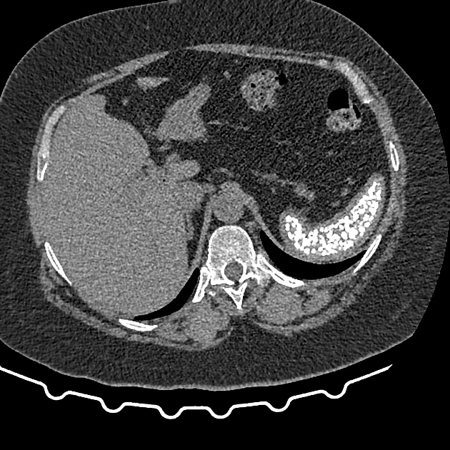
Increased splenic density can be due to a number of processes. The density may be due to calcification (most common) or other compounds (iron, Thorotrast), and can be seen (often incidentally) on abdominal radiographs and CT. On CT the usual splenic attenuation is 35-55 HU or ~10 HU 6 lower than the liver.
Pathology
Etiology
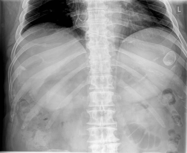
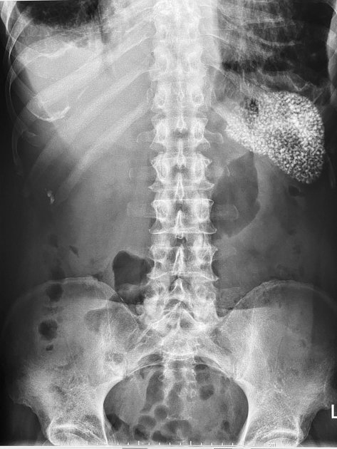
Splenic calcification
-
localized splenic calcification
- hemangiomata
- splenic infarcts 1
- cysts
- hydatid: common
- congenital, epidermoid and dermoid cysts: rarely calcify 5
- splenic hematoma 4, e.g. post traumatic
-
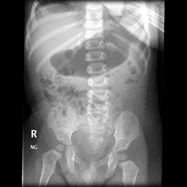
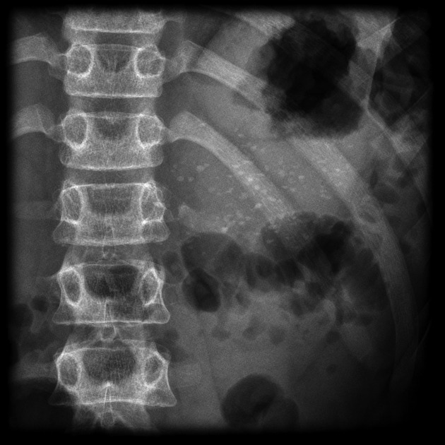
multiple focal calcifications
- granulomas 1,2: typically measure 1-10 mm in size
- Thorotrast accumulation (thorotrastosis)
See splenic calcification article for more information.
Diffuse hyperdensity
- lymphoma
- sickle cell disease
- iron deposition
- sickle cell anemia: up to 5% of cases the spleen will be increased in density, either homogeneously or with a coarse granular appearance 4 due to iron within siderotic nodules
- hemochromatosis 4,5
- Fanconi's anemia
- Thorotrast accumulation (thorotrastosis) 4
- amiodarone toxicity 7
Splenic capsule calcification
Deposition of calcium salts in on the surface of the spleen is associated with perisplenitis, secondary to peritonitis.
Differential diagnosis
-
splenic artery
- atherosclerosis of the splenic artery is very common, and may be discontinuous and partially overly the spleen 1
- splenic artery aneurysms may have calcified walls 3
On plain radiograph, consider other causes of abdominal calcification:
- pancreatic calcification as seen in chronic pancreatitis
- adrenal calcification
- costochondral calcifications
- nephrolithiasis


 Unable to process the form. Check for errors and try again.