
After normal myelination in utero, myelination of the neonatal brain is far from complete. The first myelination is seen as early as the 16th week of gestation, in the column of Burdach, but only really takes off from the 24th week 1. It does not reach maturity until 2 years or so. It correlates very closely to developmental milestones 3. The progression of myelination is predictable and abides by a few simple general rules; myelination progresses from:
central to peripheral
caudal to rostral
dorsal to ventral
sensory then motor
T1 myelination milestones
Typical T1 myelination milestones include 6:
-
term birth:
dorsal brainstem
posterior limb internal capsule
perirolandic gyri
-
3-4 months:
ventral brainstem
anterior limb internal capsule
splenium of corpus callosum
central, posterior corona radiata
-
6 months:
cerebellar white matter
genu of corpus callosum
parietal, occipital white matter
-
12 months:
posterior fossa (~adult)
most of corona radiata
posterior subcortical white matter
-
18 months:
all white matter except temporal, frontal U-fibers
-
24 months:
anterior temporal, frontal U-fibers
T2 myelination milestones
Typical T2 myelination milestones include 6:
-
term birth:
dorsal brainstem
partial posterior limb internal capsule
perirolandic gyri
-
3-4 months:
posterior limb internal capsule
-
6 months:
ventral brainstem
anterior limb internal capsule
splenium of corpus callosum
occipital white matter
-
12 months:
most of corona radiata
posterior subcortical white matter
-
18 months:
all white matter except temporal, frontal U-fibers, occipital radiations
-
24 months:
anterior temporal, frontal U-fibers
White matter myelination by location
White matter myelination also varies by location 7:
centrum semiovale: T1 at 2-4 months, T2 at 7-11 months
-
occipital white matter:
deep: T1 at 3-5 months, T2 at 9-14 months
subcortical: T1 at 4-7 months, T2 at 11-15 months
-
mid-frontal white matter:
deep: T1 at 3-6 months, T2 at 11-16 months
subcortical: T1 at 7-11 months, T2 at 14-18 months
-
anterior frontal white matter:
deep: T1 at 5-8 months, T2 at 12-18 months
subcortical: T1 at 10-15 months, T2 at 24-30 months
Radiographic features
CT
Unmyelinated white matter is hypodense compared to normal white matter and grey matter.
MRI
-
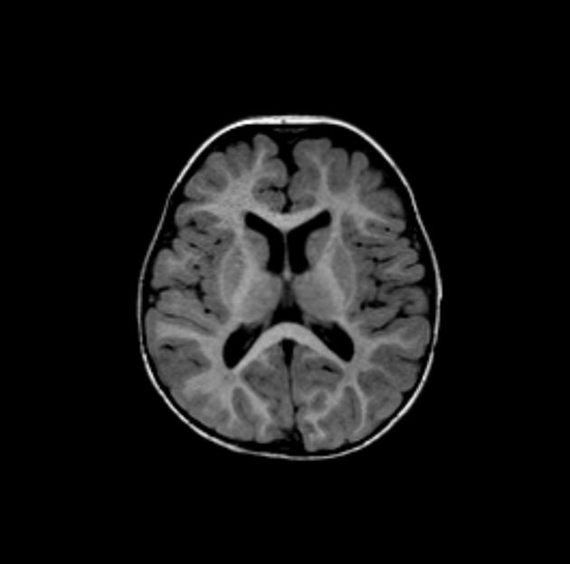
T1
most sensitive sequence in children <1 year of age 1
myelination represented by T1 hyperintensity
-
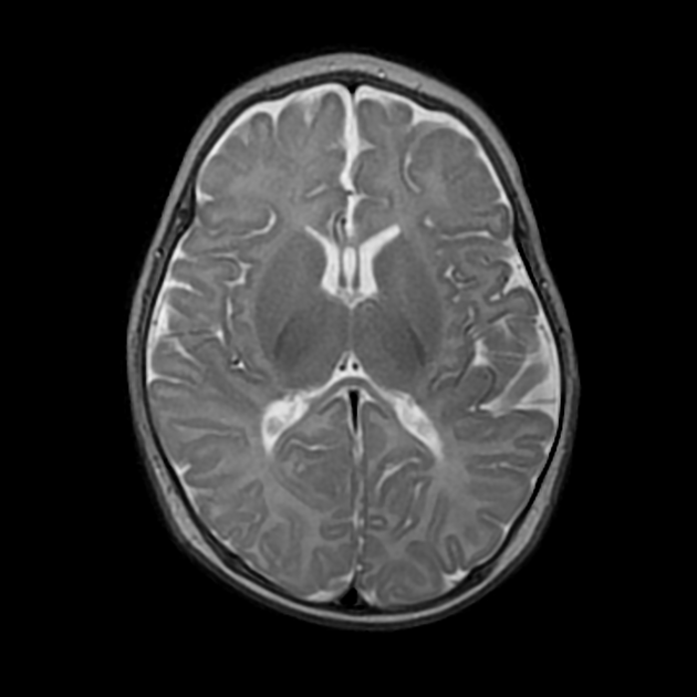
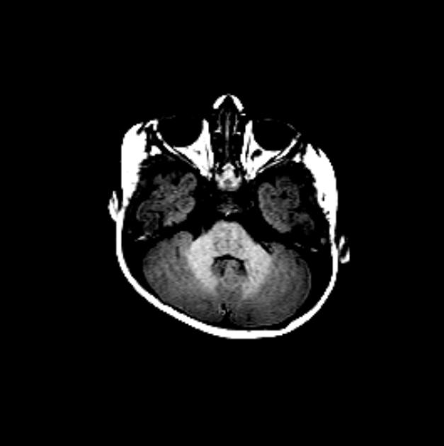
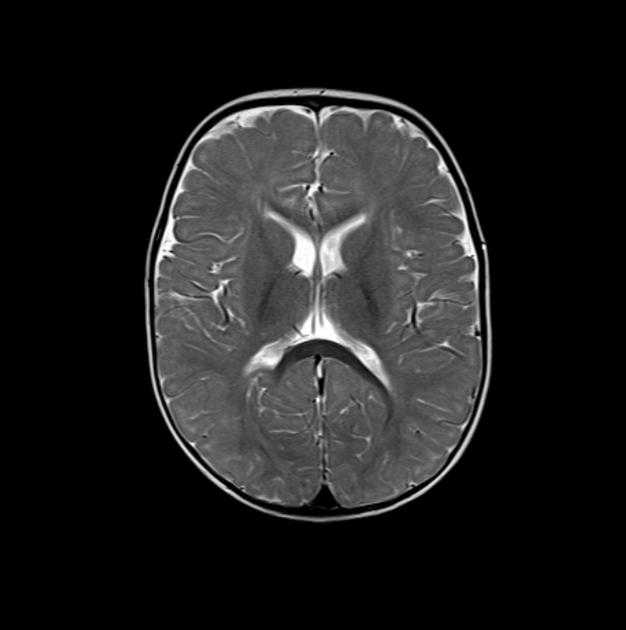
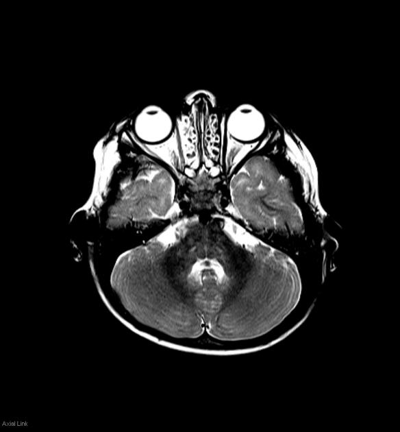
T2
most sensitive sequence in children between the age of 1 and 2 demonstrating a gradual shift from hyper- to hypo-intense relative to grey matter
the only area to remain hyperintense after the age of 2 years, and often for quite some time, is the peritrigonal region 4 which is called terminal zones of myelination
differentiation between terminal zones and PVL requires detection of normally myelinated white matter between the patchy hyperintense T2 signal and the lateral ventricle
-
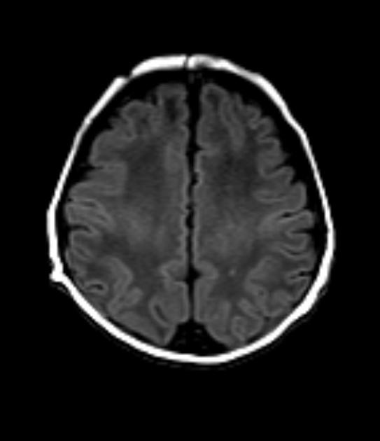
FLAIR
unsurprisingly follows the same pattern as T2 but lags behind somewhat. The exception is deep cerebral white matter, which begins as heterogeneously hypointense during the first few months of life.
this area then joins the remainder of white matter as hyperintense before finally once more becoming hypointense in the second year of life 2
-
proton density:
PD weighted images are useful in distinguishing gliosis from hypomyelination
In the acute setting, DWI is more sensitive than either T1 or T2.
Fractional anisotropy (FA) increases with brain maturation (diffusion is restricted perpendicular to the direction of axons, thus the increase in DWI signal in large axon bundles running through the slice, e.g posterior limb of internal capsule).
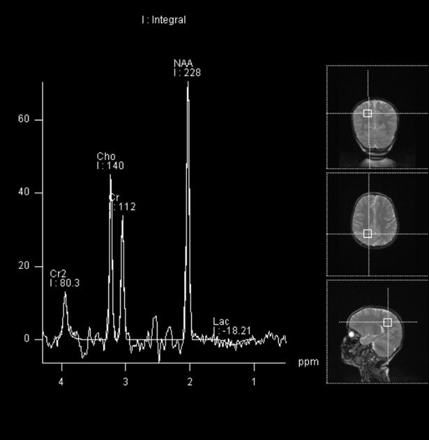
MR spectroscopy demonstrates elevated myoinositol and choline in neonates which gradually declines. Myoinositol decreases to adult values by end of first year and choline by 2-3 years. NAA increases with myelination (in the first year of life).


 Unable to process the form. Check for errors and try again.