
Esophageal intramural pseudodiverticulosis is an uncommon condition in which there are numerous small outpouchings within the esophageal wall.
On this page:
Epidemiology
It is a rare condition, found in <1% of esophagograms. It may occur at any age, but is more common between 50 and 70 years. There is a slight male predominance 2.
Associations
esophageal strictures: present in 90% of patients 1
Pathology
Intramural pseudodiverticula represent dilated excretory ducts of the deep esophageal mucosal glands 1.
Radiographic features
Fluoroscopy
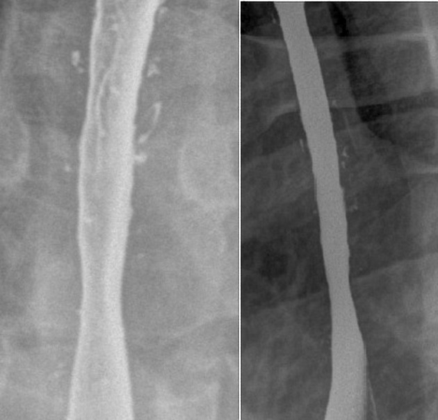
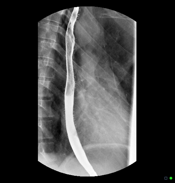
Barium swallow examination is the study of choice, as the ductal orifices may be too small to be seen on endoscopy. Pseudodiverticula are better seen with a single contrast examination than with a double contrast, thin barium examination 1,2.
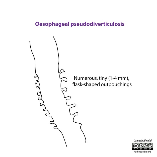
numerous, tiny (1-4 mm), flask-shaped outpouchings
may be diffusely distributed or clustered
clustering may occur next to peptic strictures
viewed in profile, often appear “floating” next to the esophageal wall, as the channel to the lumen is imperceptible
viewed en face, may appear as ulcers 3
intramural tracking may sometimes be seen bridging two or more pseudodiverticula 2
CT
In the proper clinical context, CT can confirm esophageal pseudodiverticulosis, particularly when other imaging modalities are inconclusive. The findings include:
marked thickening of the esophageal wall
diffuse irregularity of the esophageal lumen
intramural gas collections 7
Treatment and prognosis
The treatment of esophageal pseudodiverticulosis is dependent on symptoms displayed and accompanying conditions 5. Around 10% of patients do no require treatment 6. The use of proton pump inhibitors can help relieve symptoms of esophagitis. If esophageal strictures are present, the use of endoscopic dilatation helps improve treatment response 5.
Pseudodiverticular rupture with resultant mediastinitis has been reported but is very rare 4.


 Unable to process the form. Check for errors and try again.