Partial anomalous pulmonary venous return (PAPVR), also known as partial anomalous pulmonary venous connection (PAPVC), is a rare congenital cardiovascular condition in which some of the pulmonary veins, but not all, drain into the right heart or systemic venous system, rather than in the left atrium.
On this page:
Epidemiology
The overall prevalence of PAPVR is 0.4-0.7% 9.
Clinical presentation
Patients with large shunts may present with symptoms of dyspnea, chest pain and palpitations, signs like tachycardia and murmur can be encountered. Cases of secondary pulmonary arterial hypertension have been reported 6,7.
Pathology
Classification
Four types of PAPVR have been described:
-
supra-cardiac
right superior vena cava (most common)
brachiocephalic veins (=innominate veins)
-
cardiac
right atrium
coronary sinus
-
infracardiac
hepatic veins
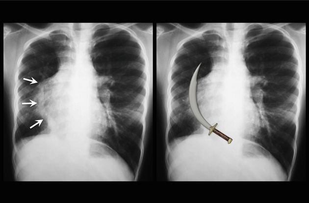
inferior vena cava (Scimitar syndrome)
-
mixed
a combination of two or more of the above anomalies
Left-sided PAPVR has been reported to be found more often in adults, whereas right-sided PAPVR is reported more commonly in children 3. It is unclear if this is because of a higher proportion of symptomatic manifestations of the latter. The left upper lobe vein anomaly is thought to be the most common.
Other rare types
Associations
in ~40% of patients with right-sided PAPVR, an atrial septal defect is seen 3
more rarely it is seen with ostium primum defect, a subtype of atrioventricular defects
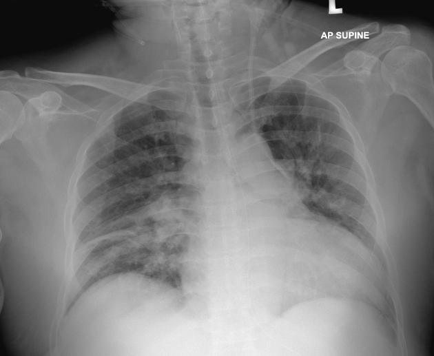
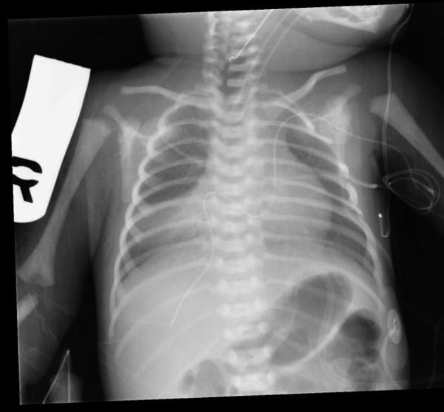
Radiographic features
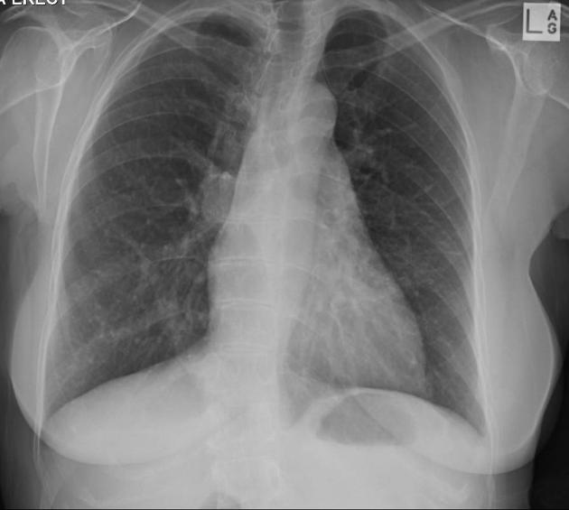
Plain radiograph
Chest radiographic features are particular to each subtype of PAPVR. The abnormal vein is rarely identified, except in cases of Scimitar syndrome. Pulmonary venous congestion can be seen if the venous drainage is obstructed.
Cardiomegaly can also be seen if significant abnormal intracardiac venous drainage occurs.
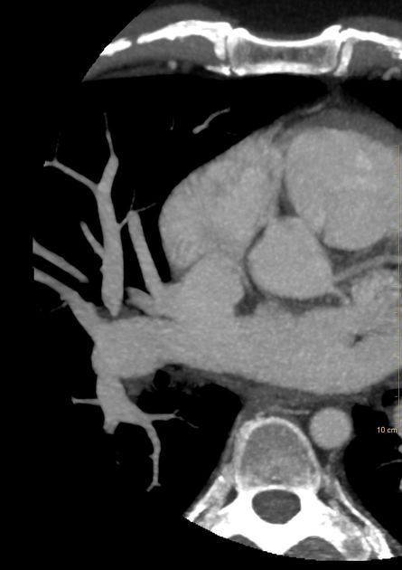
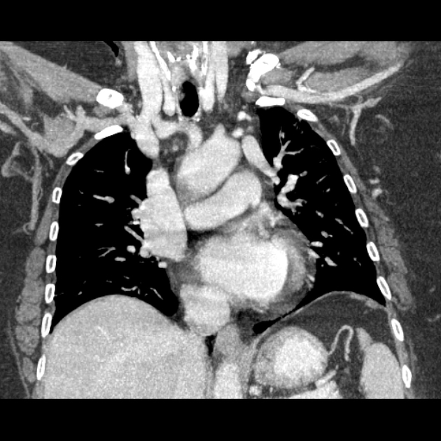
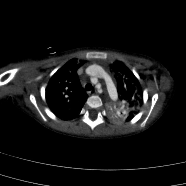
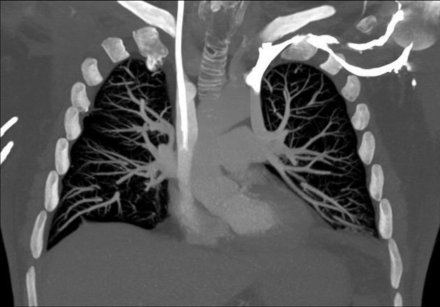
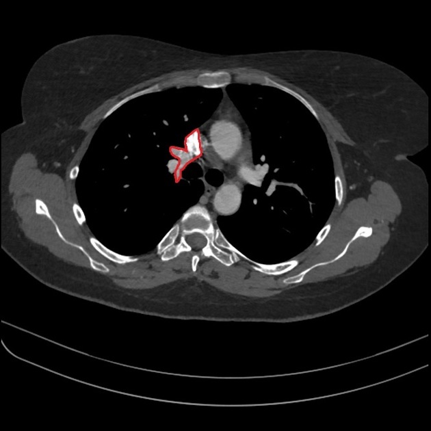
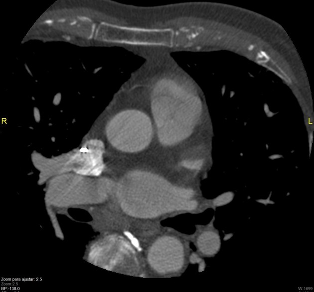
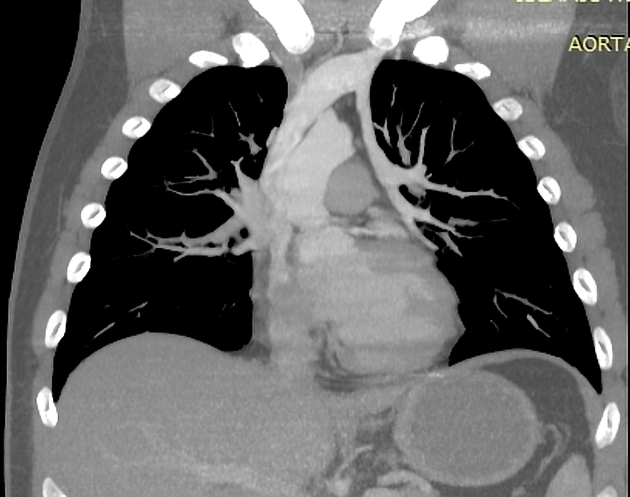
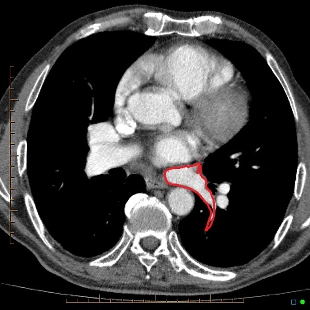
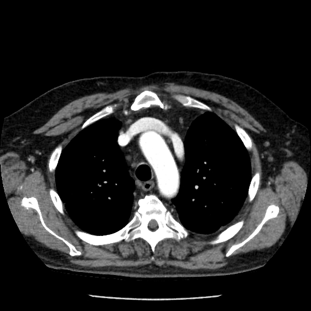
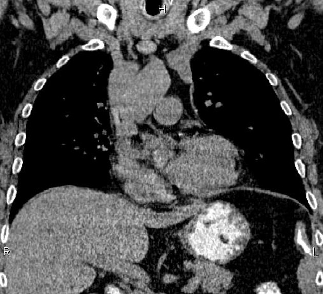
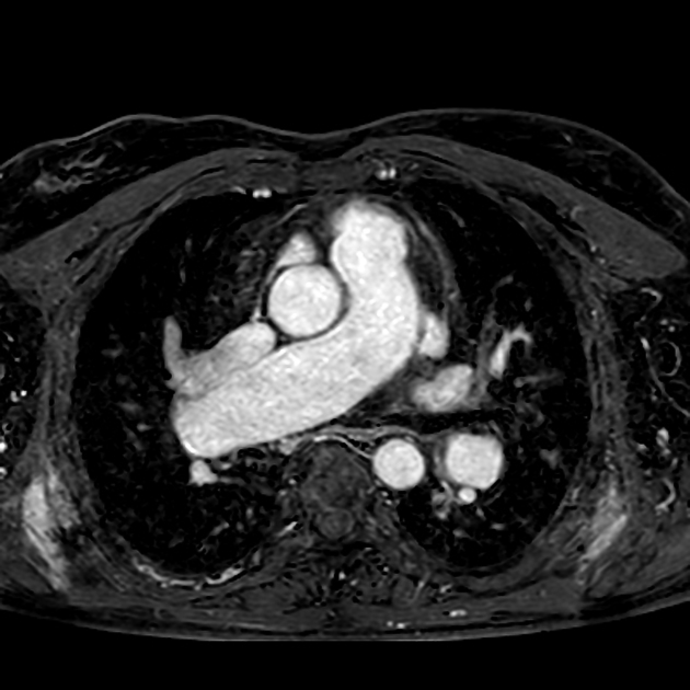
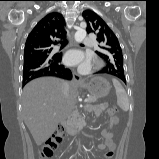
CT
Utilization of contrast-enhanced studies with MDCT technology enables both detection and characterization of the anomalies. It is considered the imaging modality of choice 3,4.
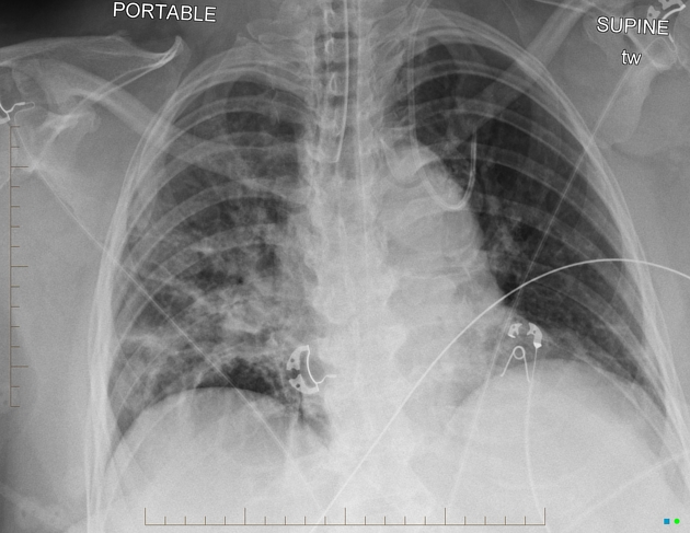
Treatment and prognosis
Therapeutic options include surgical repair with ASD patching, intracardiac baffle, anomalous vein anastomosis, systemic vein translocation and Warden procedure inter alia.
Differential diagnosis
Imaging differential considerations include:


 Unable to process the form. Check for errors and try again.