
Patent foramen ovale
Updates to Article Attributes
A patentPatent foramen ovale (PFO) is are a type of atrial septal defect in which there is channel-like communication between the atria through an unfused fossa ovalis.
AnatomyGross anatomy
The foramen ovale in the interatrial septum normally develops into the fossa ovalis when the flaps of the atrial septa primum and secundum normally fuse during development. The foramen ovale remains patent if there is incompletely fusion. This variant occurs in 25-33% of adults1,2. The prevalence may decrease with advancing age.
Patent foramen ovales/ovalia are subdivided into:
- probe-patent foramen ovale
- patent foramen ovale
A "probe patent" foramen ovale is defined as a defect in the fossa ovalis that would be revealed with instrument probing.
Pathology
A patent foramen ovale has been associated with paradoxical embolism and ischemic stroke because of the potential for a venous thromboembolism to pass from the right atrium to the systemic circulation, bypassing the lungs.
PFOs are small enough to be hemodynamically insignificant.
Radiographic features
Transesophageal echocardiography
- may be detected on a "bubble study" (IV injection of agitated saline)
. The; the contrast material appears in the left atrium before the normal timeto
Cardiac CT
- abnormal communication of contrast material between the atria through a channel-like tunnel in the interatrial septum. A channel-like tunnel alone is a normal variant of the fossa ovalis, and is not diagnostic.
Contrast-enhanced cardiac MRI
- not a first-line study, but may be diagnosed visual assessment or computation of signal–time curves in the pulmonary vein and the left atrium3
.
A patent foramen ovale can be differentiated from an atrial septal defect because a PFO takes a tunneled intraseptal course, or with the presence of a flap valve on the left atrial side of the foramen2.
Pathology
A patent foramen ovale has been associated with paradoxical embolism and ischemic stroke because of the potential for a venous thromboembolism to pass from the right atrium to the systemic circulation, bypassing the lungs.
PFOs are small enough to be hemodynamically insignificant.
TreatmentTreatment and prognosis
Closure devices, both surgically open and percutaneous, have been developed and are currently implemented in some centers for PFO.
-<p>A <strong>patent foramen ovale</strong> (<strong>PFO</strong>) is a type of <a href="/articles/atrial-septal-defect-2">atrial septal defect</a> in which there is channel-like communication between the atria through an unfused fossa ovalis.</p><h4>Anatomy</h4><p>The foramen ovale in the interatrial septum normally develops into the fossa ovalis when the flaps of the atrial septa primum and secundum normally fuse during development. The foramen ovale remains patent if there is incompletely fusion. This variant occurs in 25-33% of adults<sup>1,2</sup>. The prevalence may decrease with advancing age.</p><p>Patent foramen ovales/ovalia are subdivided into:</p><ul>- +<p><strong>Patent foramen ovale</strong> (<strong>PFO</strong>) are a type of <a href="/articles/atrial-septal-defect-2">atrial septal defect</a> in which there is channel-like communication between the atria through an unfused fossa ovalis.</p><h4>Gross anatomy</h4><p>The foramen ovale in the interatrial septum normally develops into the fossa ovalis when the flaps of the atrial septa primum and secundum normally fuse during development. The foramen ovale remains patent if there is incompletely fusion. This variant occurs in 25-33% of adults<sup>1,2</sup>. The prevalence may decrease with advancing age.</p><p>Patent foramen ovales/ovalia are subdivided into:</p><ul>
-</ul><p>A "probe patent" foramen ovale is defined as a defect in the fossa ovalis that would be revealed with instrument probing.</p><h4>Radiographic features</h4><h5>Transesophageal echocardiography</h5><ul><li>may be detected on a "bubble study" (IV injection of agitated saline). The contrast material appears in the left atrium before the normal time to </li></ul><h5>Cardiac CT</h5><ul><li>abnormal communication of contrast material between the atria through a channel-like tunnel in the interatrial septum. A channel-like tunnel alone is a normal variant of the fossa ovalis, and is not diagnostic.</li></ul><h5>Contrast-enhanced cardiac MRI</h5><ul><li>not a first-line study, but may be diagnosed visual assessment or computation of signal–time curves in the pulmonary vein and the left atrium<sup>3</sup>.</li></ul><p>A patent foramen ovale can be differentiated from an atrial septal defect because a PFO takes a tunneled intraseptal course, or with the presence of a flap valve on the left atrial side of the foramen<sup>2</sup>. </p><h4>Pathology</h4><p>A patent foramen ovale has been associated with <strong>paradoxical embolism</strong> and ischemic stroke because of the potential for a venous thromboembolism to pass from the right atrium to the systemic circulation, bypassing the lungs.</p><p>PFOs are small enough to be hemodynamically insignificant.</p><h4>Treatment</h4><p>Closure devices, both surgically open and percutaneous, have been developed and are currently implemented in some centers for PFO.</p><p> </p>- +</ul><p>A "probe patent" foramen ovale is defined as a defect in the fossa ovalis that would be revealed with instrument probing.</p><h4>Pathology</h4><p>A patent foramen ovale has been associated with <a href="/articles/paradoxical-embolism">paradoxical embolism</a> and <a href="/articles/ischaemic-stroke">ischemic stroke</a> because of the potential for a <a href="/articles/venous-thromboembolism">venous thromboembolism</a> to pass from the right atrium to the systemic circulation, bypassing the lungs.</p><p>PFOs are small enough to be hemodynamically insignificant.</p><h4>Radiographic features</h4><h5>Transesophageal echocardiography</h5><ul><li>may be detected on a "bubble study" (IV injection of agitated saline); the contrast material appears in the left atrium before the normal time</li></ul><h5>Cardiac CT</h5><ul><li>abnormal communication of contrast material between the atria through a channel-like tunnel in the interatrial septum. A channel-like tunnel alone is a normal variant of the fossa ovalis, and is not diagnostic.</li></ul><h5>Contrast-enhanced cardiac MRI</h5><ul><li>not a first-line study, but may be diagnosed visual assessment or computation of signal–time curves in the pulmonary vein and the left atrium <sup>3</sup>
- +</li></ul><p>A patent foramen ovale can be differentiated from an atrial septal defect because a PFO takes a tunneled intraseptal course, or with the presence of a flap valve on the left atrial side of the foramen <sup>2</sup>. </p><h4>Treatment and prognosis</h4><p>Closure devices, both surgically open and percutaneous, have been developed and are currently implemented in some centers for PFO.</p>
Sections changed:
- Anatomy
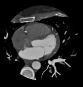
Image 1 CT (C+ arterial phase) ( create )


 Unable to process the form. Check for errors and try again.