
Portal venous phase
Citation, DOI, disclosures and article data
Citation:
Baba Y, Murphy A, Haouimi A, et al. Portal venous phase. Reference article, Radiopaedia.org (Accessed on 27 Mar 2025) https://doi.org/10.53347/rID-91786
rID:
91786
Article created:
Disclosures:
At the time the article was created Yahya Baba had no recorded disclosures.
View Yahya Baba's current disclosures
Last revised:
Disclosures:
At the time the article was last revised Andrew Murphy had no financial relationships to ineligible companies to disclose.
View Andrew Murphy's current disclosures
Revisions:
14 times, by
4 contributors -
see full revision history and disclosures
Systems:
Sections:
Synonyms:
- Hepatic phase
- Late portal phase
- Portal venous phase (PVP)
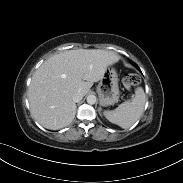
The portal venous phase, also known as the late portal phase or hepatic phase, is a contrast-enhanced CT or MRI series that has the following characteristics:
- liver parenchyma is at its peak enhancement with a density >110 HU (an increase of at least 50 HU from the unenhanced baseline)1,2
- portal vein and hepatic veins are completely enhanced
On this page:
Technique
The acquisition time depends on the intravenous device (central or peripheral), the concentration of the contrast medium, and the injection rate.
-
time from injection through an upper extremity vein: 70-90 seconds 3
- 70 seconds at an injection rate of 4 ml/s 4
- time from bolus tracking: 50-70 seconds
Clinical use
The portal venous phase offers the best hepatic enhancement for the detection of:
See also
References
- 1. Heiken JP, Brink JA, McClennan BL, Sagel SS, Crowe TM, Gaines MV. Dynamic incremental CT: effect of volume and concentration of contrast material and patient weight on hepatic enhancement. (1995) Radiology. 195 (2): 353-7. doi:10.1148/radiology.195.2.7724752 - Pubmed
- 2. Fujigai T, Kumano S, Okada M, Hyodo T, Imaoka I, Yagyu Y, Ashikaga R, Ishii K, Murakami T. Optimal dose of contrast medium for depiction of hypervascular HCC on dynamic MDCT. (2012) European journal of radiology. 81 (11): 2978-83. doi:10.1016/j.ejrad.2012.01.016 - Pubmed
- 3.Mitsuzaki K, Yamashita Y, Ogata I, Nishiharu T, Urata J, Takahashi M. Multiple-phase helical CT of the liver for detecting small hepatomas in patients with liver cirrhosis: contrast-injection protocol and optimal timing. (1996) AJR. American journal of roentgenology. 167 (3): 753-7. doi:10.2214/ajr.167.3.8751695 - Pubmed
- 4. Monzawa S, Ichikawa T, Nakajima H, Kitanaka Y, Omata K, Araki T. Dynamic CT for detecting small hepatocellular carcinoma: usefulness of delayed phase imaging. (2007) AJR. American journal of roentgenology. 188 (1): 147-53. doi:10.2214/AJR.05.0512 - Pubmed
Incoming Links
Related articles: Computed tomography
- computed tomography in practice
-
computed tomography overview
- iodinated contrast media
- CT IV contrast media administration
-
CT artifacts
- patient-based artifacts
- physics-based artifacts
- hardware-based artifacts
- ring artifact
- tube arcing
- out of field artifact
- air bubble artifact
- helical and multichannel artifacts
- CT technology
-
generations of CT scanners
- helical CT scanning
- step and shoot scanning
- ultra-high-resolution CT (UHRCT)
- CT x-ray tube
- CT fluoroscopy
- cone-beam CT
-
generations of CT scanners
- dual-energy CT
- CT image reconstruction
- CT image quality
- CT dose
-
CT protocols
- composite
- head & neck
- chest
- abdomen and pelvis
- CT abdomen-pelvis (protocol)
- CT abdominal aorta
- CT adrenals (protocol)
- CT cholangiography (protocol)
- CT colonography (protocol)
- CT enteroclysis (protocol)
- CT enterography (protocol)
- CT gastrography (protocol)
- CT kidneys, ureters and bladder (protocol)
- CT urography (protocol)
- CT Renal mass (protocol)
- CT angiography of the splanchnic vessels (protocol)
- CT renal split bolus
- CT pancreas (protocol)
- liver


 Unable to process the form. Check for errors and try again.