Cardiac gating (CT)
Citation, DOI & article data
- Gated CT
- ECG gated cardiac CT
Cardiac gating or ECG gated angiography in CT is an acquisition technique that triggers a scan during a specific portion of the cardiac cycle. Often this technique is conveyed to obtain high-quality scans void of pulsation artefact.
On this page:
Technique
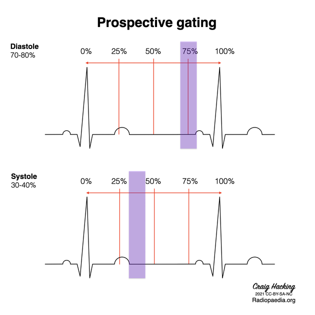
Via the attachment of ECG leads, cardiac gating aims to find a consistent portion of the R-R interval (measured as a percentage), most commonly at the mid to end-diastolic phase (around 70% of the R-R interval) 1, where movement is at its least.
For higher heart rates (>80bpm) end-systole acquisition (30-35% of R-R interval, or 350ms) 2 is often utilized.
Gated protocols in cardiac CT
There are a number of different protocols based on the patient presentation that can be broken into prospective and retrospective based:
- prospective
- stable sinus rhythm and a heart rate of fewer than 60 beats a minute
- stable sinus rhythm and a heart rate of fewer than 70 beats a minute
- retrospective
- atrial fibrillation/arrhythmia
Prospectively ECG-gated cCTA protocols
Prospective protocols mean a predetermined portion of the R-R interval is selected for scanning. It is not a functional study, however useful to reduce motion artifact.
Prospective ECG gated (step and shoot)
This technique (also called step-and-shoot mode) is based on the principle that several images acquired at different heart phases and anatomic regions of the heart can be combined for reconstruction of the whole heart.
High-pitch spiral acquisition
Dual-source CT scanners allow for a gapless acquisition with a pitch of up to 3.4 which cannot be achieved with conventional single-source CT scanners. A high-pitch spiral acquisition can be performed in less than one second and thus information from a single heartbeat can be generated. In combination with iterative reconstruction techniques, high-pitch spiral acquisition allows for cardiac CT with sub-milliSievert doses.
Limitations of prospectively ECG-gated protocols
It should be noted that all prospectively ECG-gated protocols are more susceptible to deterioration of image quality due to motion artifacts. Therefore, when choosing such a protocol, the patient's heart rate should ideally be below 65 beats per minute (bpm). In high-pitch spiral acquisition, the heart rate should even be below 60 bpm. Therefore, the cost of significant dose reduction is the limited patient population that such protocols can be applied to.
Retrospectively ECG-gated cCTA protocols
A retrospective protocol means after detection of the heart rhythm, the scan covers the whole heart during multiple cardiac cycles.
This is an established and robust technique which works as a standard spiral CT acquisition. Therefore, information from different phases of the cardiac cycle is gained and can be used. This is especially useful for cine-sequences which show the heart motion throughout the cardiac cycle. Currently, such cine sequences can only be generated from retrospectively ECG-gated cCTA data.
Although several dose-saving techniques for retrospectively ECG-gated cCTA exist (e.g. tube modulation), these protocols still result in a higher radiation dose than newly introduced prospectively ECG-gated protocols. They do however retain their clinical relevance in patients with arrhythmia and for volumetry and functional analysis.
Specific artery selection
Each artery will suffer from motion artefact at different phases, the following intervals are the best ‘windows’ to prevent movement for different arteries 1
-
right coronary artery
- 40% of R-R interval
-
left anterior descending
- 60-70% of R-R interval
-
left circumflex artery
- 50-60% of R-R interval
References
- 1. Tae-Hwan Lim. Practical Textbook of Cardiac CT and MRI. (2015) ISBN: 9783642363979
- 2. Haruhiko Machida, Isao Tanaka, Rika Fukui, Yun Shen, Takuya Ishikawa, Etsuko Tate, Eiko Ueno. Current and Novel Imaging Techniques in Coronary CT. (2015) RadioGraphics. doi:10.1148/rg.2015140181
Related articles: Imaging in practice
- imaging in practice
-
general radiography (adult)
- portable radiography
- chest radiography
- abdominal radiography
-
upper limb radiography
-
shoulder girdle radiography
- scapula series
-
shoulder series
- shoulder (AP view)
- shoulder (internal rotation view)
- shoulder (external rotation view)
- shoulder (superior-inferior axial view)
- shoulder (inferior-superior axial)
- shoulder (West Point view)
- shoulder (Velpeau view)
- shoulder (modified trauma axial view)
- shoulder (supine lateral view)
- shoulder (modified transthoracic supine lateral)
- shoulder (lateral scapula view)
- shoulder (AP glenoid view)
- shoulder (Garth view)
- shoulder (outlet view)
- shoulder (Stryker notch view)
- acromioclavicular joint series
-
clavicle series
- clavicle (AP view)
- clavicle (AP cephalic view)
- clavicle (oblique view)
- sternoclavicular joint series
- arm and forearm radiography
- wrist and hand radiography
- wrist series
- scaphoid series
- hand series
- thumb series
- fingers series
- rheumatology hands series
- bone age (radiograph)
-
shoulder girdle radiography
-
lower limb radiography
- pelvic girdle radiography
- thigh and leg radiography
- ankle and foot radiography
- skull radiography
-
paranasal sinus and facial bone radiography
- facial bones
- nasal bones
- zygomatic arches
- paranasal sinuses
- temporal bones
- dental radiography
- orthopantomography
- mandible
- mandible (PA view)
- mandible (PA axial view)
- mandible (AP axial view)
- mandible (oblique view)
- temporomandibular joints
- temporomandibular joint (AP axial view)
- temporomandibular joint (axiolateral view)
- temporomandibular joint (axiolateral oblique view)
-
spinal radiography
- cervical spine series
-
thoracic spine series
- thoracic spine (AP view)
- thoracic spine (lateral view)
- thoracic spine (oblique view)
- lumbar spine series
- sacrococcygeal radiography
- scoliosis radiography
-
pediatric radiography
- radiographic positioning terminology
- systematic radiographic technical evaluation (mnemonic)
- pediatric immobilization
- foreign body ingestion series (pediatric)
- foreign body inhalation series (pediatric)
- shunt series
- chest radiograph (pediatric)
- abdomen radiograph (pediatric)
-
upper limb radiography (pediatric)
- shoulder series (pediatric)
- pediatric shoulder (AP view)
- pediatric shoulder (lateral view)
- clavicle series (pediatric)
- pediatric clavicle (AP view)
- pediatric clavicle (axial view)
- humerus series (pediatric)
- elbow series (pediatric)
- forearm series (pediatric)
- wrist series (pediatric)
- scaphoid series (pediatric)
- hand series (pediatric)
- thumb series (pediatric)
- finger series (pediatric)
- shoulder series (pediatric)
- lower limb radiography (pediatric)
- pelvis radiograph (pediatric)
- femur series (pediatric)
- pediatric femur (AP view)
- pediatric femur (lateral view)
- leg length view
- knee series (pediatric)
- pediatric knee (AP view)
- pediatric knee (lateral view)
- tibia fibula series (pediatric)
- pediatric tibia fibula (AP view)
- pediatric tibia fibula (lateral view)
- pediatric tibia fibula (oblique view)
- ankle series (pediatric)
- pediatric ankle (AP view)
- pediatric ankle (mortise view)
- pediatric ankle (lateral view)
- foot series (pediatric)
- pediatric foot (DP view)
- pediatric foot (oblique view)
- pediatric foot (lateral view)
- skull radiography (pediatric)
- spine radiography (pediatric)
-
skeletal survey
-
skeletal survey (non-accidental injury)
- torso
- pediatric chest (AP erect view)
- pediatric chest (oblique ribs view)
- pediatric abdomen (AP supine view)
- pediatric spine (whole lateral view)
- upper limb (both sides)
- lower limb (both sides)
- pediatric femur (AP view)
- pediatric knee (lateral view)
- pediatric tibia fibula (AP view)
- pediatric ankle (lateral view)
- pediatric foot (DP view)
- torso
-
skeletal survey (non-accidental injury)
-
CT
- iodinated contrast media
- CT IV contrast media administration
-
CT protocol
- composite
- whole-body CT (protocol)
- CT Chest abdomen-pelvis (protocol)
- CT NCAP (neck, chest, abdomen and pelvis)
- head & neck
- chest
- abdomen and pelvis
- CT abdomen-pelvis (protocol)
- CT abdominal aorta
- CT adrenals (protocol)
- CT cholangiography (protocol)
- CT colonography (protocol)
-
CT enteroclysis (protocol)
-
CT enterography (protocol)
- CT gastrography (protocol)
- CT kidneys, ureters and bladder (protocol)
- CT urography (protocol)
- CT Renal mass (protocol)
- CT angiography of the splanchnic vessels (protocol)
- CT renal split bolus
- CT pancreas (protocol)
- liver
- composite
- barium studies
-
MRI
- cine imaging
-
brain
- screen protocol
- stereotaxis protocol
- tumor protocol
- stroke protocol
- infection protocol
- trauma protocol
- demyelination protocol
- epilepsy protocol
- neurodegenerative protocol
- trigeminal neuralgia protocol
- posterior fossa protocol
- temporal bone/IAM/CPA protocol
- pineal and tectal plate protocol
- pituitary gland protocol
- CSF flow
- angiographic protocols
- MRA
- circle of Willis (COW)
- carotid-vertebral system
- MRV
- MRA
- head and neck
- orbits protocol
- sinonasal tract protocol
- salivary glands protocol
- oropharynx and oral cavity protocol
- suprahyoid neck protocol
- infrahyoid neck protocol
- chest
- abdomen and pelvis
- abdomen
- liver and biliary tree
- liver protocol
- MRCP: cholangiopancreatography
- pancreas protocol
- elastography
- anus and rectum
- uterus and ovaries
- urological
- prostate cancer protocol
- bladder cancer protocol
- kidneys protocol
- musculoskeletal
-
ultrasound
- ultrasound signs
-
obstetric ultrasound
- other
- placenta
-
second trimester
- fetal biometry
- fetal morphology assessment
-
soft markers
- nuchal fold thickness
- ventriculomegaly
- absent nasal bone
- echogenic intracardiac focus
- choroid plexus cysts
- echogenic bowel
- aberrant right subclavian artery
- amnioreduction
- echogenic fetal bowel
- umbilical artery Doppler assessment
- chorionic villus sampling (CVS) and amniocentesis
- nuchal translucency
- failed early pregnancy
- subchorionic hematoma
- multiple gestations
- ectopic pregnancy
-
first trimester and early pregnancy
- gestational sac
- yolk sac
- embryo/fetus
- amnion
- chorion
- Beta-hCG levels
- gynecologic ultrasound
- vascular ultrasound
- carotids
- extremities
- mesenteric vessels
- other
- breast ultrasound
-
musculoskeletal ultrasound
- technique/artifacts
- ultrasound of arthropathies
- skin/soft tissue ultrasound
- lipoma
- parasitic infection
- other
- pediatric musculoskeletal ultrasound
- ankle/foot ultrasound
-
knee ultrasound
- Baker cyst (popliteal cyst)
- infrapatellar bursitis
- hip ultrasound
- hand ultrasound
- wrist ultrasound
- elbow ultrasound
- shoulder ultrasound
- liver ultrasound
- hyperechoic liver lesion
- sonographic halo sign
- ultrasound appearances of liver metastases
- periportal hyperechogenicity
- periportal hypoechogenicity
- generalized increase in hepatic echogenicity
- generalized reduced hepatic echogenicity
- coarsened hepatic echotexture
- starry sky appearance (ultrasound)
- normal hepatic vein Doppler
- hepatic arterial resistive index
- gallbladder ultrasound
- pancreatic ultrasound
- gastrointestinal ultrasound
- renal ultrasound
- bladder ultrasound
-
testicular and scrotal ultrasound
- other
- epididymis
- paratesticular lesions
- bilateral testicular lesion
-
unilateral testicular lesion
- testicular torsion
- orchitis
- testicular rupture
-
germ cell tumor of the testis
- testicular seminoma
-
non seminomatous germ cell tumors
- mixed germ cell tumor
- yolk sac tumor (endodermal sinus tumor)
- embryonal cell carcinoma
- choriocarcinoma
- testicular teratoma
- testicular epidermoid (teratoma with ectodermal elements only)
- burned out testis tumor
- sex cord / stromal tumors of the testis
- prostate ultrasound
- neck and thyroid ultrasound
- echocardiography
- speckle tracking echocardiography
- fetal echocardiography
- contrast-enhanced echocardiography
- epicardial echocardiography
- three dimensional (3D) echocardiography
- transesophageal echocardiography (TEE)
- transthoracic echocardiography (TTE)
- left ventricular systolic and diastolic function
- structure and morphology
- systolic function
- diastolic function
- right ventricular assessment
- right and left atria
- valvular structure and function
- mitral valve
- aortic valve
- pulmonic valve
- tricuspid valve
- hemodynamics
- pericardium
- cardiomyopathies
- congenital heart disease
- great vessels
- pediatric ultrasound
- ultrasound interventions
- ultrasound-guided biopsy
- ultrasound-guided percutaneous drainage
- ultrasound-guided musculoskeletal interventions
- joint injection
- nerve blocks
- ultrasound-guided intravenous cannulation
- contrast-enhanced ultrasound
- physics and imaging modes
- grey-scale (B-mode)
- motion mode (M-mode)
- color flow Doppler (CFD)
-
spectral Doppler
- pulsed wave Doppler (PWD)
- continuous wave Doppler (CWD)
- superb microvascular imaging (SMI)
- tissue Doppler imaging (TDI)
- nuclear medicine
-
radiation therapy
- external beam radiation therapy (EBRT)
- sealed source radiation therapy (brachytherapy)
- unsealed source radiation therapy
-
interventional
- procedure overview
- neck
- thyroid gland
- breast
- chest
- hepatobiliary
- splenic interventions
- gastrointestinal
- urogenital
- bladder
- kidney
- prostate
- musculoskeletal
-
arthrogram
- MR arthrogram
- CT arthrogram
- anesthetic arthrogram
- bone biopsy (CT-guided)
-
arthrogram
- upper limb
- lower limb
-
hip
- hip joint injection (technique)
- greater trochanteric bursa injection
- gluteus minimus/medius tendon calcific tendinopathy barbotage
- iliopsoas tendon bursa injection
- lateral cutaneous femoral nerve of the thigh injection
- piriformis injection
- common hamstrings origin injections
-
knee
- common peroneal (fibular) nerve injection
- knee joint injection
- patella tendon microtenotomy
- quadriceps tendon microtenotomy
- tibial nerve injection
-
ankle
- achilles hydrodilation & microtenotomy
- ankle joint injection
- extensor tendon sheath injection
- flexor tendon sheath injection
- os trigonum injection
- peroneal tendon sheath injection
- subtalar joint injection
- tibial nerve injection
-
foot
- calcaneocuboid joint injection
- metatarsophalangeal joint (MTPJ) injection
- naviculocuneiform joint injection
- plantar fascia microtenotomy
- subtalar joint injection
- talonavicular joint injection
- tarsometatarsal joint (TMTJ) injection
-
hip
- spine
- breast imaging
-
mammography
- breast screening
- breast imaging and the technologist
- forbidden (check) areas in mammography
-
mammography views
- craniocaudal view
- mediolateral oblique view
- additional (supplementary) views
- true lateral view
- lateromedial oblique view
- late mediolateral view
- step oblique views
- spot view
- double spot compression view
- magnification view
- exaggerated craniocaudal (axillary) view
- cleavage view
- tangential views
- caudocranial view
- bullseye CC view
- rolled CC view
- elevated craniocaudal projection
- caudal cranial projection
- 20° oblique projection
- inferomedial superolateral oblique projection
- Eklund technique
- normal breast imaging examples
-
mammography
- digital breast tomosynthesis
- breast ultrasound
- breast ductography
- breast MRI
- breast morphology
- breast intervention