
Pulmonary alveolar microlithiasis
Updates to Article Attributes
Pulmonary alveolar microlithiasis (PAM) is a rare idiopathic condition characterised by widespread intra-alveolar deposition of spherical calcium phosphate microliths (calcospherites).
Epidemiology
A slight female predilection may be present in familial form 2. Most cases are reported in Asia and Europe 9.
Clinical presentation
Often discovered incidentally on a chest radiograph. The radiographic features are frequently out of proportion to clinical symptoms 5.
Pathology
PAM is believed to be due to a mutation in SLC34A2 that causes inactivation of a sodium-dependent phosphate cotransporter, which itself is found mainly in alveolar type II cells. This cotransporter normally clears phosphate from degraded surfactant, and when inactivated there is accumulation of phosphate in the alveolus, and calcium phosphate microliths are then thought to form 9.
An autosomal recessive inheritance pattern has been proposed given familial occurrence in a majority of cases 2-3,9. Usually, there is no abnormal calciummetabolism.
Associations
Radiographic features
The pattern in children differs from that in adults 2.
Plain radiograph
Chest x-ray typically demonstrates:
- sand-like calcification distributed throughout the lungs
- bilateral distribution with middle to lower zone predilection 2,3
- black pleural lines may be evident 2
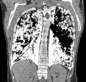
CT
HRCT better shows numerous sand-like calcifications throughout the lungs with subpleural and peribronchial distribution (typically ~1 mm) 8. Additional accompanying HRCT features include
- crazy paving pattern 1,5
- calcified interlobular septa 1
- small subpleural cysts / emphysema 1
- black pleura sign
- ground glass opacities: tends to be more common in children 2
Treatment and prognosis
No known treatment is available. Overall prognosis is good but occasionally slow progression can result in end-stage lung fibrosis requiring lung transplantation.
Differential diagnosis
Some consider the radiographic appearance to be pathognomonic 3.
Differential for fine granular densities includes
- silicosis
- healed varicella pneumonia
- idiopathic pulmonary haemosiderosis: tend to be large nodular opacities; no calcification 1
- pulmonary stannosis: deposition of tin dust
- pulmonary baritosis: deposition of barium dust
- talcosis
- mitral stenosis: tends to give small foci of pulmonary ossification
See also
- differential for small hyperdense pulmonary nodules
- differential for miliary opacities
- differential for crazy paving pattern
- differential for ground glass densities
-<a title="idiopathic pulmonary haemosiderosis" href="/articles/idiopathic-pulmonary-haemosiderosis">idiopathic pulmonary haemosiderosis</a>: tend to be large nodular opacities; no calcification <sup>1</sup>- +<a href="/articles/idiopathic-pulmonary-haemosiderosis">idiopathic pulmonary haemosiderosis</a>: tend to be large nodular opacities; no calcification <sup>1</sup>
Image 8 CT (non-contrast) ( create )


 Unable to process the form. Check for errors and try again.