
Radiographic positioning terminology is used routinely to describe the position of the patient for taking various radiographs. Standard nomenclature is employed with respect to the anatomic position.
Terminology
Basic terms of relations
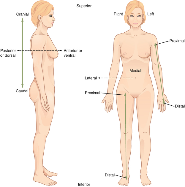
anterior is towards the front of the body (Latin: before)
posterior is towards the back of the body (Latin: after)
superior is towards the top of the body (Latin: above)
inferior is towards the bottom of the body (Latin: below)
-
medial is towards the midline (Latin: middle)
compared with median which is in the midline rather than towards the midline
lateral is away from the midline (Latin: side)
proximal is towards the center of the body (Latin: near)
distal is away from the center of the body (Latin: far)
superficial is towards the surface of the body
deep is away from the surface of the body
ipsilateral is on the same side of the body
contralateral is on the opposite side of the body
Planes
-
axial plane (transverse or transaxial plane): horizontal plane perpendicular to the long axis of the body
divides the body into superior and inferior parts
-
sagittal plane: vertical plane parallel to the median plane (or midsagittal plane)
divides the body into right and left halves
-
coronal plane: vertical plane perpendicular to the median plane
divides the body into anterior and posterior parts
Body positions
erect: either standing or sitting
supine (decubitus): lying on back
semi-erect: partially sat up
Trendelenburg position: the patient is supine (on an inclined radiographic table) with the head lower than the feet
prone: lying face-down
-
lateral: side touches the cassette
right lateral: right side touches the cassette
left lateral: left side touches the cassette
-
lateral decubitus: lying on one side, cassette is anterior/posterior
right lateral decubitus: lying on right side
left lateral decubitus: lying left side
Movement
flexion: decrease in the angle of the joint
extension: increase in the angle of the joint
abduction: movement of limb away from midline
adduction: movement of limb towards the midline
pronation: movement of hand and forearm to bring the palm facing posterior
supination: movement of hand and forearm to bring the palm facing anterior
circumduction: circular movement of a joint using a combination of flexion, abduction, extension and adduction such that the distal limb describes a circle
opposition: thumb brought to oppose another digit
reposition: thumb repositioned back to the anatomic position
elevation: movement of the scapular superiorly
depression: movement of the scapular inferiorly
eversion: movement of the sole of the foot away from the median plane
inversion: movement of the sole of the foot towards from the median plane
protrusion: movement of the mandible, lips or tongue anteriorly
retraction: movement of the mandible, lips or tongue posteriorly
Projections
Depending on patient presentation, a single view or orthogonal projections comprising of the list projections below may be performed to visualize the region of interest.
anteroposterior (AP): central ray passes, perpendicular to the coronal plane, from anterior to posterior
-
posteroanterior (PA): central ray passes, perpendicular to the coronal plane, from posterior to anterior
depending on the anatomic segment to radiograph, synonyms can be used, for example: occipito-frontal (skull); dorso-ventral (thorax); dorso-palmar (hand)
-
lateral: central ray, perpendicular to the sagittal plane and parallel to the coronal plane, passes from one side of body to the other
horizontal beam lateral (HBL): lateral view obtained with central ray projecting horizontally; generally the patient will be supine (e.g. post trauma)
oblique: central ray passes through the body/body part through a plane which is at an angle to the transverse plane/coronal plane
-
axial: central ray passes through (or parallel) to the long axis of the body
in some cases, however, the central ray runs through (or parallel) to the long axis of the skeletal segment studied (for example, the axial view of the calcaneus)
tangential: central ray is directed along the surface of the bone to image adjacent soft tissue e.g. tangential skull soft tissue x-ray for foreign body
en-face: used in imaging foreign bodies, the central ray follows the trajectory of the foreign body to discern soft tissue versus bony involvement


 Unable to process the form. Check for errors and try again.