
Spinal ependymomas are the most common spinal cord tumor overall, seen both in adult and pediatric populations.
This article specifically relates to spinal cord ependymomas. For a discussion of posterior fossa ependymomas and for a general discussion of the pathology refer to the main article: ependymoma. Additionally, due to different imaging features and demographics, supratentorial ependymomas and myxopapillary ependymomas are also discussed separately.
On this page:
Epidemiology
Spinal ependymomas are the most common intramedullary neoplasm in adults, comprising 60% of all glial spinal cord tumors 7. They are the second most common intramedullary neoplasm in the pediatric population, representing 30% of pediatric intramedullary spinal neoplasms 6.
Peak incidence is in the fourth decade, with 39 years being the mean age at presentation. Males are more commonly affected than females.
There is an increased coincidence with neurofibromatosis type 2.
Clinical presentation
Clinical presentation is similar to that of other intramedullary spinal tumors, with pain, weakness, and sensory changes common.
The frequent presentation of sensory symptoms may be explained by the proximity of these centrally located tumors to the spinothalamic tracts. Dominant motor symptoms are commonly associated with very large ependymomas 5.
Pathology
Ependymomas were thought to arise from ependymal cells lining the central canal of the spinal cord, but it has become evident that they actually arise from progenitor cells or radial glia-like stem cells 10.
Microscopic appearance
On histologic evaluation, uniform moderately hyperchromatic nuclei are typically observed. Perivascular pseudorosettes are the classical finding.
Genetics
In a small subgroup of patients, MYCN-amplification is identified. This is a distinct tumor and is now recognized as a separate entity in the 5th edition (2021) of the WHO classification of CNS tumors 10.
Grading
The vast majority of spinal ependymomas are WHO grade 2, with grade 3 tumors being rare.
Spinal ependymomas with MYCN-amplification are high-grade aggressive tumors, however, they are yet to be given a formal grade in the 5th edition (2021) of the WHO classification of CNS tumors 10.
Radiographic features
Ependymomas can occur anywhere along the spinal cord, however, the cervical cord is the most common site (44%). An additional 23% occur within the cervical cord and extend into the upper thoracic cord, and 26% occur in the thoracic cord alone 3.
Plain radiograph
Plain film features that may be seen with a spinal ependymoma include:
CT
CT may demonstrate:
non-specific canal widening
iso to slightly hyper-attenuating compared with normal spinal cord
intense enhancement with iodinated contrast
large lesions may cause scalloping of the posterior vertebral bodies and neural exit foraminal enlargement
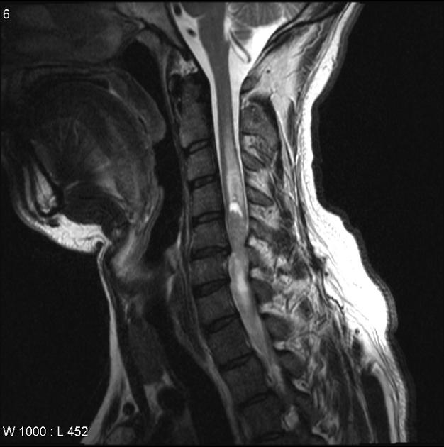
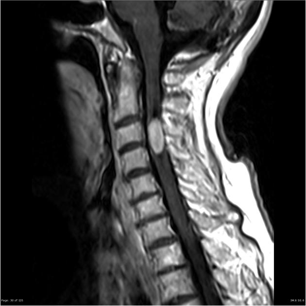
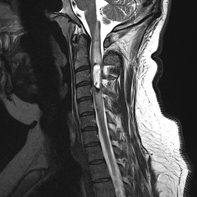
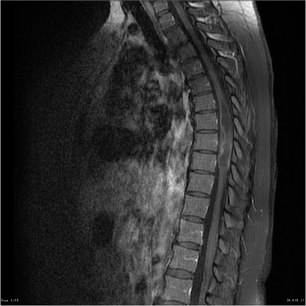
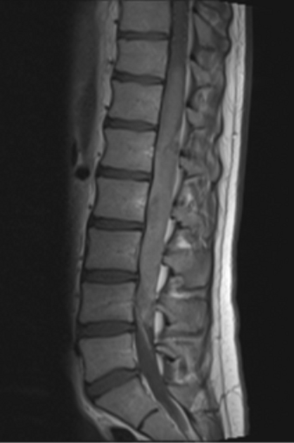
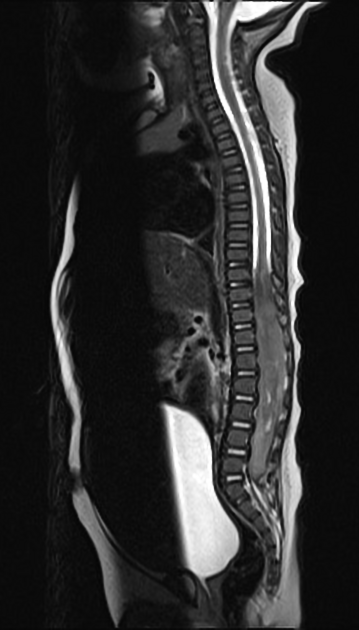
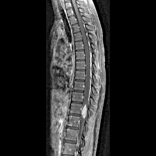
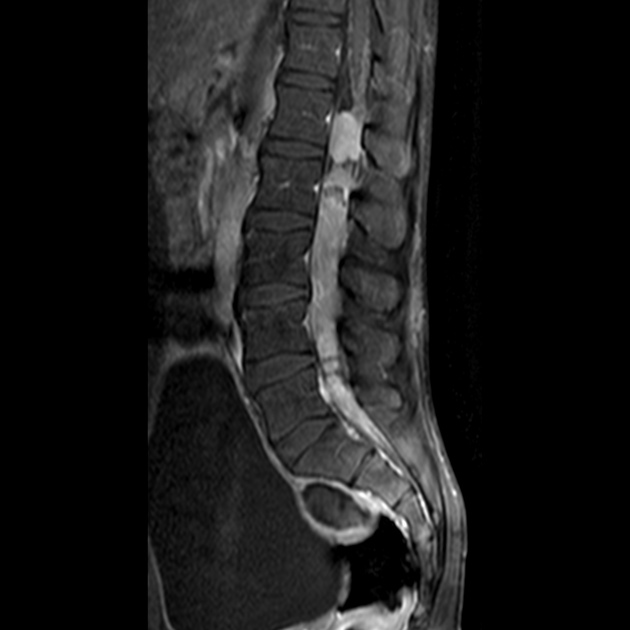
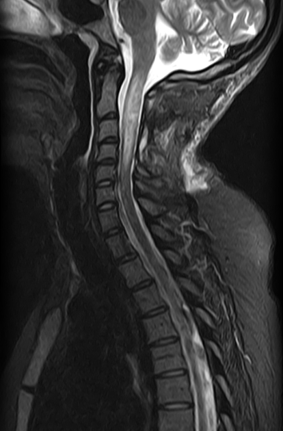
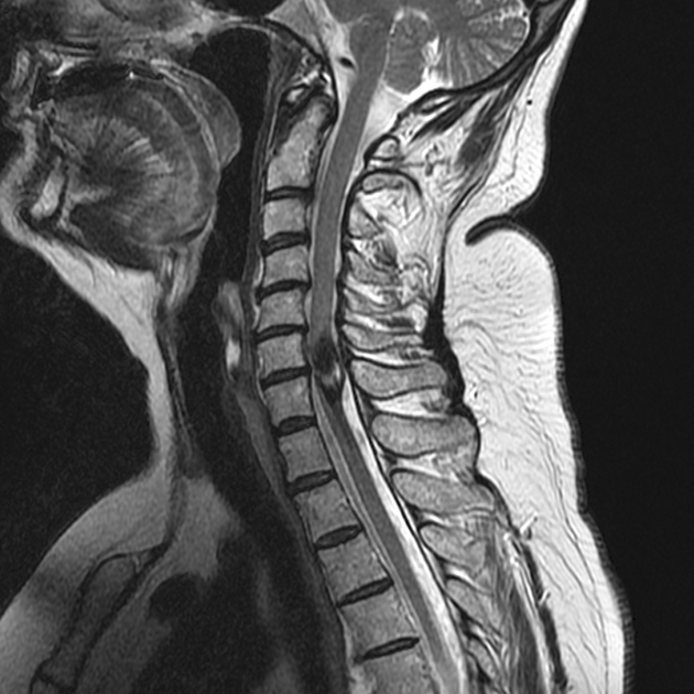
MRI
MRI is the modality of choice for evaluating suspected spinal cord tumors. Features include:
widened spinal cord (as ependymomas arise from ependymal cells lining the central canal, they tend to occupy the central portion of the spinal cord and cause symmetric cord expansion)
although unencapsulated, they are well-circumscribed
tumoral cysts are present in 22%; non-tumoral cysts are present in 62% 5
syringohydromyelia occurs in 9-50% of cases
in contrast to intracranial ependymomas, calcification is uncommon
average length of four vertebral body segments 10
Signal characteristics
T1: most are isointense to hypointense; mixed-signal lesions are seen if cyst formation, tumor necrosis or hemorrhage has occurred
-
T2: hyperintense
peritumoural edema is seen in 60% of cases
associated hemorrhage leads to the “cap sign” (a hypointense hemosiderin rim on T2 weighted images) in 20-33% of cases 5. The cap sign is suggestive of but not pathognomonic for ependymoma as it may also be seen in hemangioblastomas and paragangliomas
T1 C+ (Gd): virtually all enhance strongly, somewhat inhomogeneously
Video
Treatment and prognosis
Most ependymomas are slow-growing. They tend to compress adjacent spinal cord tissue rather than infiltrate it, almost always leaving a cleavage plane between the tumor and spinal cord tissue.
A complete curative excision may be achieved in approximately 50% of cases. In those patients, the 5-year survival rate is approximately 85%. In patients who are not able to achieve complete resection, the 5-year survival rate is approximately 57%. Recurrence is rare following complete excision 6.
Although metastatic spread is rare, the most common sites for metastases include the retroperitoneum, lymph nodes, and lungs 5.
Spinal ependymomas with MYCN-amplification are, in contrast, aggressive tumors with poor prognosis and frequent early dissemination throughout the central nervous system and reoccurrence despite resection10.
Differential diagnosis
On MRI, the main differential diagnosis is an astrocytoma. Certain imaging features may help to differentiate between the two, which are covered in this article:
-
most common spinal cord tumor in children
eccentric location in the spinal canal
ill-defined
hemorrhage is uncommon
patchy irregular contrast enhancement
bone changes are infrequent
involvement of the entire cord diameter and longer cord segments favors an astrocytoma (i.e., if an intramedullary neoplasm involves the total spinal cord, it is more likely to be an astrocytoma 9 )
-
no enhancement
complete hemosiderin ring


 Unable to process the form. Check for errors and try again.