
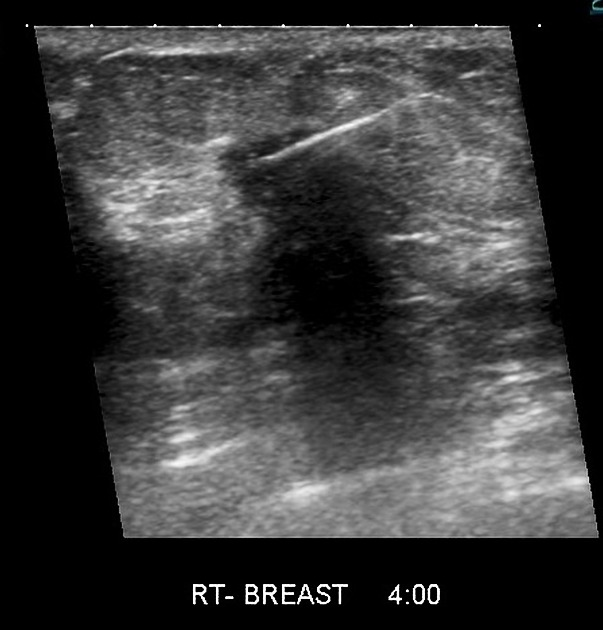
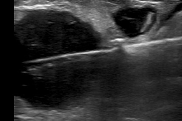
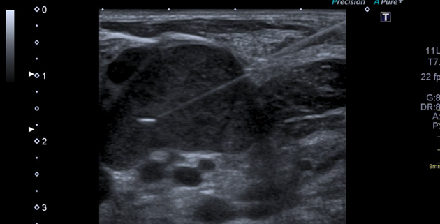
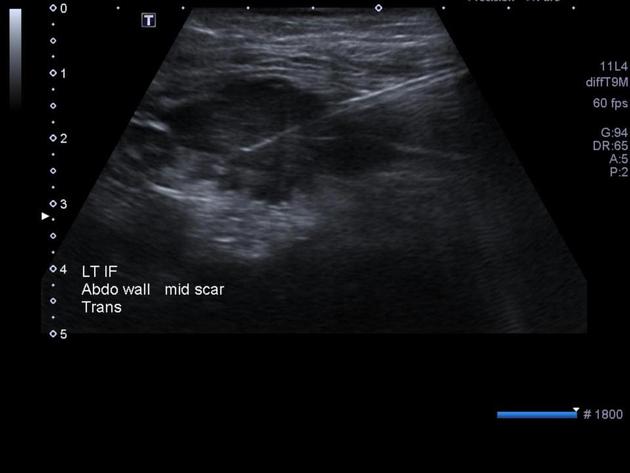
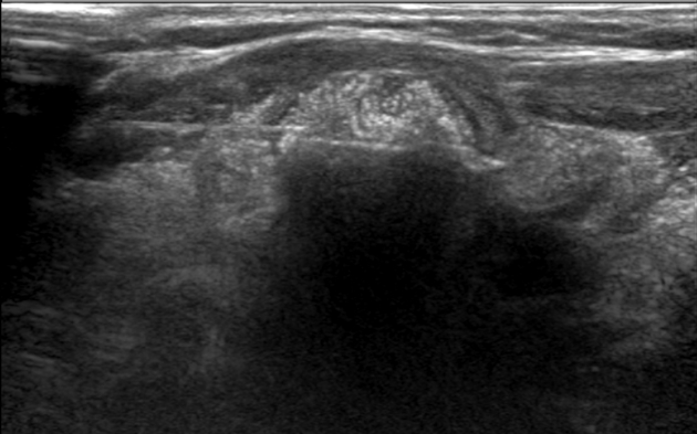
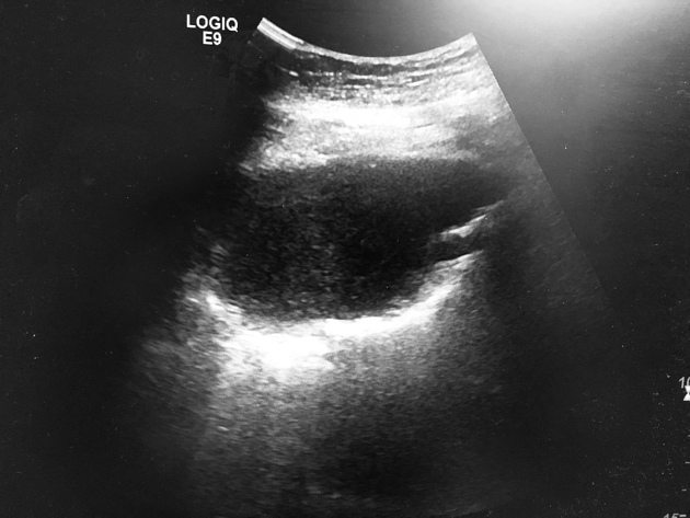
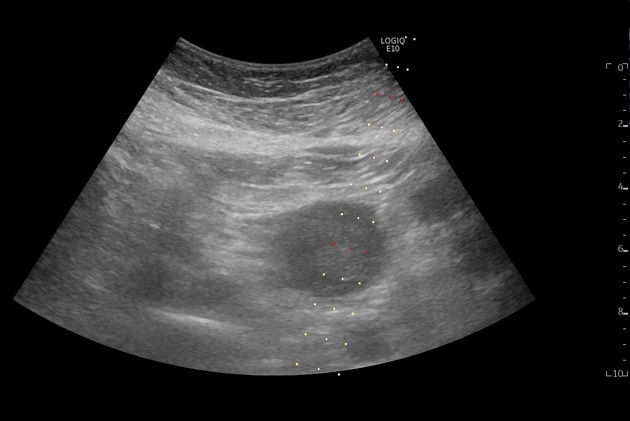
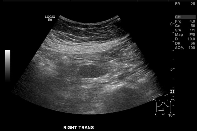
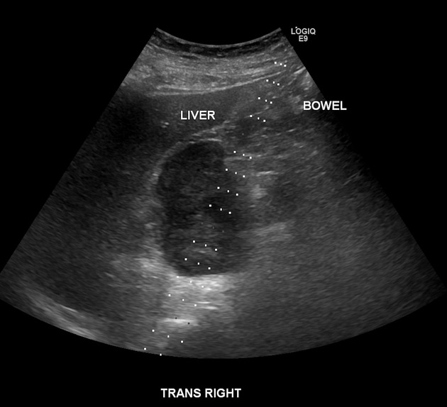
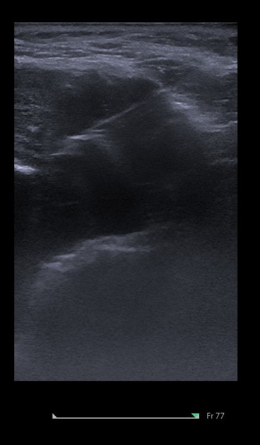
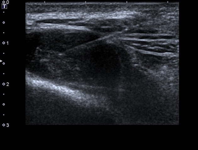
Ultrasound-guided biopsy is one form of image-guided biopsy, typically performed by a radiologist. It is the most common form of image-guided biopsy, offering convenience and real-time dynamic observation with echogenic markers on cannulae allowing for precise placement.
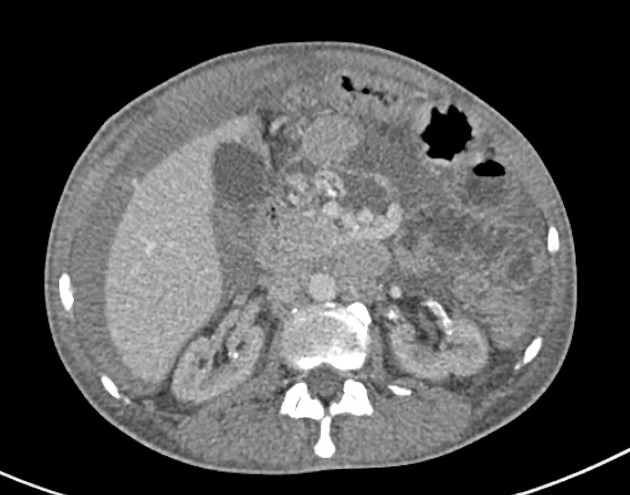
It can potentially be used to perform a biopsy of any body part, being regularly used to biopsy the kidney, liver, breast and lymph nodes. There can be some minor procedural variation in the type of biopsy performed.
Biopsies may be focal or non-focal in nature. Lesions as small as 5 mm may be biopsied, with a range of biopsy needle gauges and sizes available (from 22G to 14G). Broadly speaking needles may be single or co-axial and have 10 mm or 20 mm cutting lengths.
Most common percutaneous biopsies
Laboratory parameters for a safe procedure
Biopsies in deep structures require special attention to coagulation indices. There are widely divergent opinions about the safe values of these indices for percutaneous biopsies. The values suggested below were considered based on the literature review, whose references are cited below.
Complete blood count: platelets >50,000/mm3 (some institutions determine other values between 50,000-100,000/mm3) 1,3.
Coagulation profile: some studies showed that having a normal INR or prothrombin time is no reassurance that the patient will not bleed after the procedure 2.
international normalized ratio (INR) ≤1.5 3
normal prothrombin time (PT), partial thromboplastin time (PTT)
Contraindications
The contraindications must be considered individually in each case. Overall, the most important contraindications to percutaneous biopsies are:
uncooperative patient
lack of safe access
uncorrectable bleeding diathesis (abnormal coagulation indices)


 Unable to process the form. Check for errors and try again.