
The vertebral venous plexus is a highly anastomotic network of valveless veins running along the entire length of the vertebral column from the foramen magnum to the sacral hiatus.
Gross anatomy
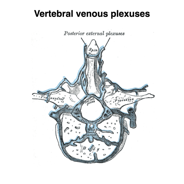
The vertebral venous plexus is comprised of three interconnected divisions:
internal vertebral venous plexus
external vertebral venous plexus
basivertebral veins
The internal vertebral venous plexuses and external vertebral venous plexuses communicate via the intervertebral veins, which run through the intervertebral foramina.
Venous return within the epidural plexus proceeds through a superior or inferior route, depending on the anatomic level. At or above L2, the plexus is drained by the lumbar veins, which join the inferior vena cava but also may continue their cephalad course into the azygos or hemiazygos system 3. Below L2, drainage is "descendant", with foraminal veins draining into the iliolumbar veins, which are tributaries of the common iliac veins 3.
Internal vertebral venous plexus
The internal vertebral venous plexuses consist of two anterior and two posterior interconnecting longitudinal vessels lying in the epidural space surrounded by a small collection of semi-liquid fat. They receive tributaries from the radicular and basivertebral veins as well as the Batson plexus.
The anterior internal vertebral venous plexus is more prominent and largely lies posterior to the vertebral bodies, while the posterior internal vertebral venous plexus largely lies anterior to the laminae.
External vertebral venous plexus
The external vertebral venous plexuses are composed of the anterior and posterior external vertebral plexuses that surround the vertebral column. In addition to the internal vertebral venous plexus, they form several connections with the azygos, lumbar and deep cervical veins.
The anterior external vertebral venous plexus lies anterior to the vertebral bodies while the posterior external vertebral venous plexus lies posterior to the vertebral arch.
Basivertebral veins
The basivertebral veins course horizontally within the vertebral bodies, receiving tributaries from numerous small venous channels. These are drained posteriorly by the anterior internal vertebral venous plexus and anteriorly by the external vertebral venous plexus.
Related pathology
As such, and owing to its unvalved nature, the vertebral venous plexus is subject to distension in cases of increased intrathoracic or intra-abdominal pressure due to ascites, pregnancy, large tumors, etc. This leads to an increased risk of trauma during needle placement into the epidural space.


 Unable to process the form. Check for errors and try again.