
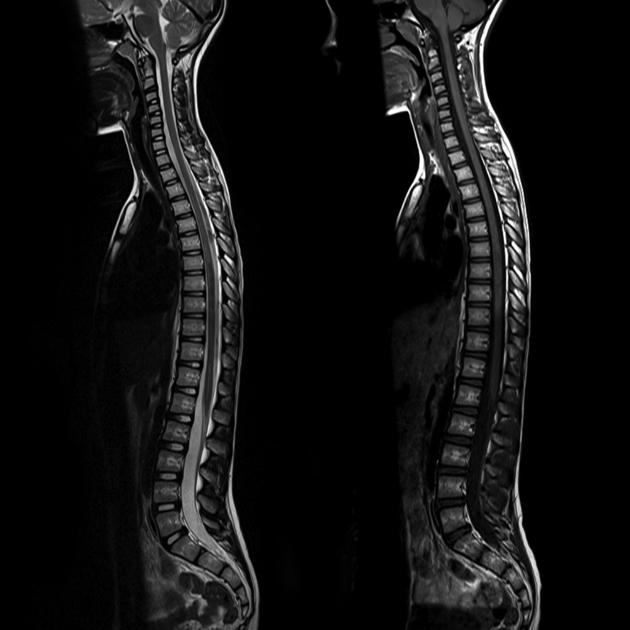
The spinal cord is the part of the central nervous system found within the vertebral column's spinal canal. The cord extends from the corticomedullary junction at the foramen magnum of the skull down to the tip of the conus medullaris within the lumbar cistern. It is lined by the spinal pia mater and contained by the other spinal meninges in the thecal sac.
On this page:
Terminology
Although the spinal cord is anatomically divided into cervical, thoracic, lumbar, sacral and coccygeal segments on the basis of segments that correspond to the emerging nerve roots, due to the disproportionate growth of the spine compared to the cord, these segments do not match adjacent vertebral bodies. In clinical radiology practice, as it is difficult to identify the spinal cord segments on imaging, the spinal cord is usually referred to relative to the spine. In other words, the cervical cord is the cord segment extending from the base of skull to the C7/T1 disc level, and the thoracic cord extends from there to the T12/L1 disc. The very tip is the conus medullaris, usually located at approximately L1/2, although this is variable. The segment of the cord between the conus medullaris and T12/L1 is rarely, if ever, referred to as the lumbar cord.
Gross anatomy
The spinal cord measures approximately 42-45 cm in length, ~1 cm in diameter and 35 g in weight 1,2,8. The caliber of the cord is not, however, uniform along its length. Rather it has two expansion corresponding to the upper limbs (cervical expansion; C3-T2) and lower limbs (lumbar expansion; T9-T12) 8.
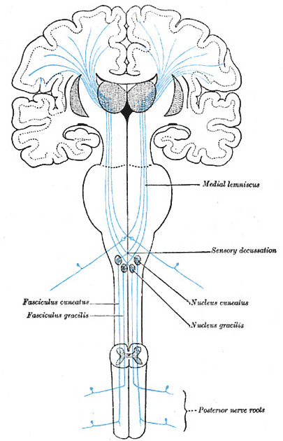
Like the brain, it is composed of grey and white matter. However, unlike the brain, the grey matter is on the internal aspect of the cord, and the white matter tracts are external. Throughout its length, paired dorsal and ventral nerve roots enter its dorsolateral and ventrolateral surface, respectively.
The spinal cord is divided into cervical, thoracic and lumbar parts and terminates at the tip of the conus medullaris at approximately the L1 vertebral body level in adults.
The spinal cord is segmented by the 31 nerve roots that emerge from it:
8 cervical
12 thoracic
5 lumbar
5 sacral
1 coccygeal
Internal structure
A transverse section of the spinal cord shows a peripheral mass of white matter surrounding an ‘H’ or butterfly-shaped central mass of grey matter with a small ependyma-lined central canal filled with CSF 2. The cord is incompletely divided into left and right halves by the posterior median sulcus (shallow) and the anterior median fissure (deep) 6.
Grey matter
The grey matter contains the cell bodies of neurons and glia and is enlarged in the cervical and lumbosacral regions to provide fibers to the large nerve plexuses. It is divided into anterior, dorsal and lateral horns and periependymal grey matter 3,5:
-
anterior horns
contain motor neurons for skeletal muscle
send efferent fibers via the ventral nerve roots
-
lateral horns
contains preganglionic sympathetic cell bodies
only present at levels which contain these cell bodies (T1 to L2/3) 7
-
dorsal horns
contain somatosensory neurons
receive primary afferents from the dorsal roots of the spinal nerves
-
periependymal grey matter
divided into ventral and dorsal grey matter commissures
White matter
The white matter contains nerve fibers or tracts and is divided into anterior, dorsal and lateral columns (also known as funiculi) as well as the anterior spinal commissure 3,5.
-
columns
anterior columns primarily contain the spinothalamic tracts which are responsible for pain, temperature, coarse (non-discriminative) touch and pressure sensations
dorsal columns contain ascending fibers which are responsible for vibration, conscious proprioception, and fine (discriminative) touch sensations
lateral columns primarily contain the corticospinal tracts which are the principal motor pathway connecting the cerebral cortex to spinal motor neurons
anterior spinal commissure is located between the posterior-most extent of the anterior median fissure anteriorly and the ventral grey matter commissure posteriorly 5.
In general, fibers found posteriorly process and relay sensory information, fibers found laterally are preganglionic visceral motor neurons and somatic motor fibers are found anteriorly 4.
Blood supply
Blood supply is discussed separately. See spinal cord blood supply.
Related pathology
-
spinal tumors and cysts
-
-
open spinal dysraphism
-
closed spinal dysraphism
-
-
traumatic
-
spinal ischemia
-
spinal vascular malformations
-
others


 Unable to process the form. Check for errors and try again.