
The atlas (plural: atlases) is the first cervical vertebra, commonly called C1. It is an atypical cervical vertebra with unique features. It articulates with the dens of the axis and the occiput, respectively allowing rotation of the head, and flexion, extension and lateral flexion of the head. Unlike the rest of the cervical vertebrae, with exception to the similarly structured axis, the anterior rami are sent posteriorly instead of anteriorly.
On this page:
Gross anatomy
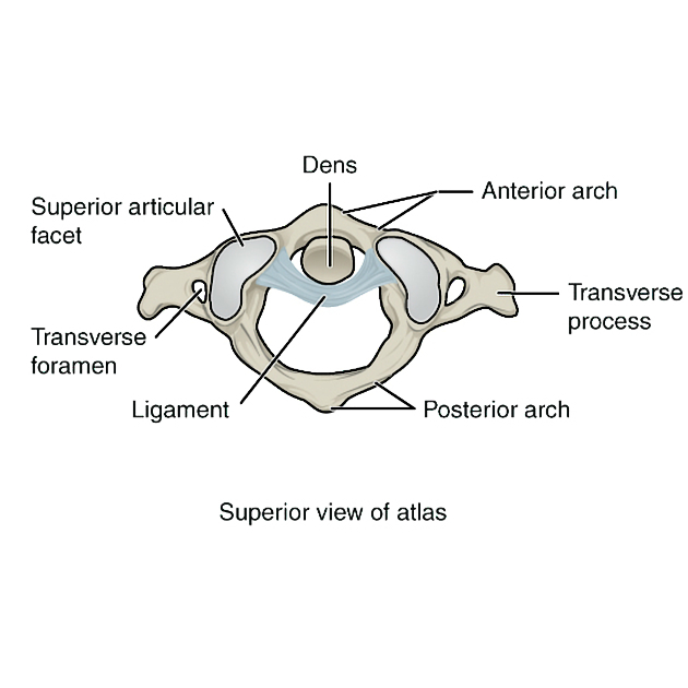
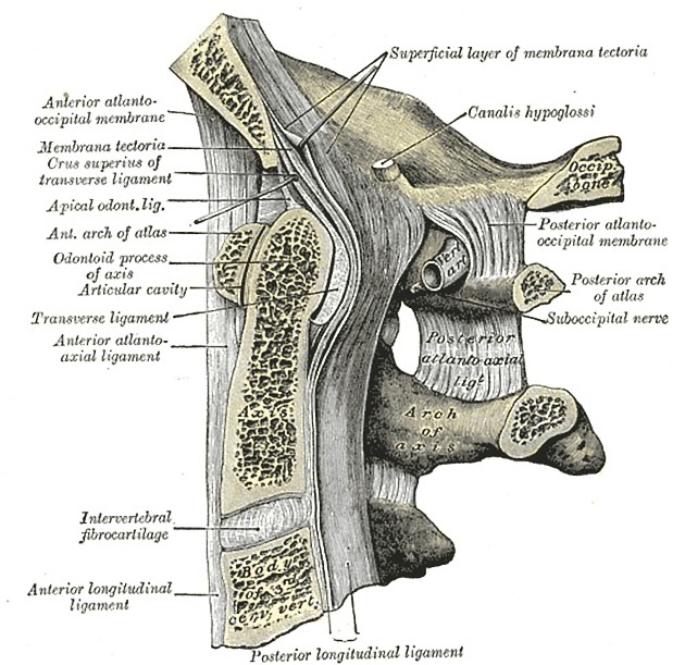
The atlas is composed of an anterior arch and a posterior arch, paired lateral masses, and paired transverse processes. It does not have a vertebral body, instead the dens of the axis sit where a centrum (body) of a typical vertebra would be. The transverse ligament holds the dens of the axis against the anterior arch of the atlas and divides its vertebral canal into two parts. The anterior 1/3 is occupied by the dens. The posterior 2/3 contains the spinal cord, which occupies 1/3 of the total vertebral canal space.
-
anterior arch
anterior tubercle: sits on the anterior aspect of the anterior arch and is the site of attachment of the anterior longitudinal ligament
posterior facet for the dens: sits on the posterior aspect of the anterior arch
upper border: attachment of the anterior atlanto-occipital membrane and lateral parts of the anterior longitudinal ligament
lower border: attachment of the anterior atlanto-axial membrane and lateral parts of the anterior longitudinal ligament
-
posterior arch
3/5 of circumference of the ring
posterior tubercle: sits on the posterior aspect of the posterior arch, is a rudimentary spinous process and attachment site for the ligamentum nuchae
superior surface: contains paired grooves for the C1 nerve and vertebral artery, sits just posterior to the lateral mass
superior border: attachment for the posterior atlanto-occipital membrane
inferior border: attachment for the posterior atlanto-axial membrane
-
lateral masses
paired, ovoid
superior articular facet: kidney-shaped, concave and articulates with the occipital bone
inferior articular facet: circular, with a flat or slightly concave surface articulating with the lateral atlantoaxial joint
medial surface: marked by vascular foramina and a tubercle for the attachment of the transverse ligament
-
transverse processes
longer than all of the transverse processes of the cervical vertebrae except C7
typically covered by costal lamella
transverse foramina: contains the vertebral arteries
anterior tubercle: sometimes present on the anterior aspect of the transverse process
Articulations
atlanto-occipital joint: hyaline-covered synovial joint between the occipital condyle and concave facet of the lateral mass of the atlas. Covered by a capsule and innervated by C1, this joint allows for flexion, extension and lateral flexion.
median atlantoaxial joint: hyaline-covered synovial joint between the dens of the axis and the posterior aspect of the anterior arch of the atlas, allowing for the rotation of the head. The dens is held in place by the transverse ligament, with a bursa between the two.
lateral atlantoaxial joint: hyaline-covered synovial joint between the inferior articular facet of the atlas and the superior articular facet of the axis which allows for the rotation of the head. A capsule innervated by the C2 nerve surrounds the joint.
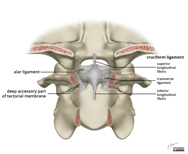
Ligamentous
transverse ligament: strong band that runs posterior to the dens of the axis, holding it in place. Each end is attached to tubercles on the anterior arch of the atlas.
atlantoaxial ligaments: attach from the lower border of the anterior arch of the atlas to front of the body of the axis. Provides tertiary support against ventral translation of the dens.
Musculotendinous
anterior atlanto-occipital membrane: attached to upper border of the anterior arch to the outer margins of foramen magnum
posterior atlanto-occipital membrane: attached to upper border of the posterior arch to the outer margins of foramen magnum. At each lateral margin, there is a gap for the passage of the C1 nerve and vertebral artery, which sometimes ossifies and becomes a foramen. Innervated by C1.
longus colli: anterior tubercle
levator scapulae: tip of the transverse processes
splenius cervicis: transverse processes
obliquus capitis superior: transverse processes
obliquus capitis inferior: transverse processes
rectus capitis anterior: base of transverse processes
rectus capitis posterior minor: tubercle on the posterior arch of the atlas
Variant anatomy
arcuate foramen: calcification of the posterior atlanto-occipital membrane
superior articular facets of the lateral masses can be divided into two parts, with the anterior part being larger and the posterior smaller
-
fusion defects
central or paramedian parts of the posterior arch can be absent and replaced by fibrous tissue
anterior arch fusion defects
split atlas - rare congenital fusion defects of both the anterior and posterior arches
fusion (with the occiput - partially or fully)
Radiographic features
Plain radiograph
Transoral AP view best appreciates the atlanto-axial joint
anterior atlanto-dental interval: <3 mm ventral translation is normal 6
sum of displacement of lateral masses of C1 compared to C2 >8 mm on transoral AP view 6
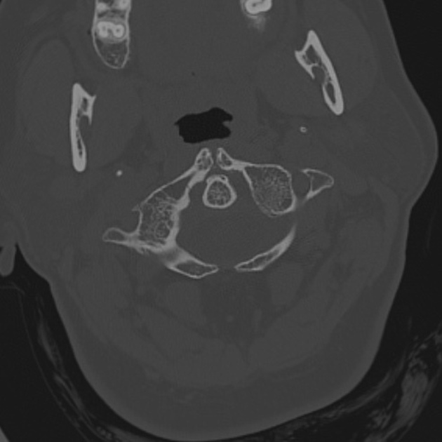
CT
used for assessment of fractures
MRI
best appreciates ligamentous disruption or avulsion
Development
Ossification starts from three centers: anterior ossification center, and paired lateral mass ossification centers. The paired lateral mass ossification centers arise in week 7 and extend to the posterior arch. Unification at the posterior arch occurs at years 3-4 and is typically direct, but can sometimes involve a third center at the posterior arch. The anterior ossification center unites with the lateral mass ossification center at years 6-8. There are sometimes two anterior ossification centers.
History and etymology
Atlas was the name of the Classical Greek god who bore the world on his shoulders 3.
Related pathology
-
Gehweiler classification of atlas fractures 6
type 1: isolated posterior arch fracture
type 2: isolated anterior arch fracture
type 3 (Jefferson fracture): anterior and posterior arch fractures
type 4: comminuted lateral mass fracture


 Unable to process the form. Check for errors and try again.