
Intervertebral discs form the articulations between adjacent vertebral bodies from C2 to S1, thus forming 23 in total (although segmentation variations are not uncommon). Together they account for ~25% of the total height of the vertebral column which decreases with age as disc height is lost through normal aging.
On this page:
Gross anatomy
The intervertebral discs are secondary cartilaginous joints, also known as symphyses 7.
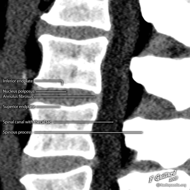
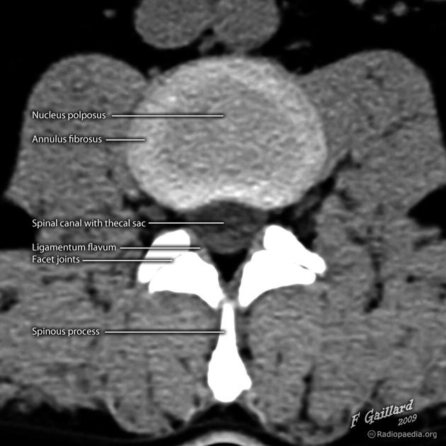
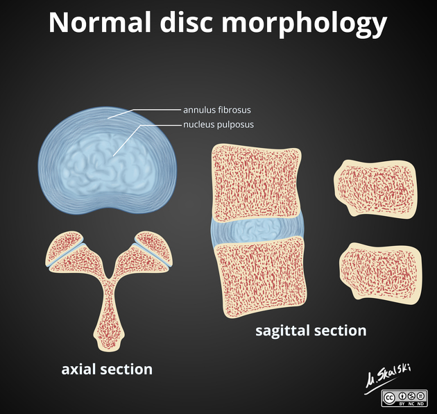
Each intervertebral disc is comprised of:
peripheral annulus fibrosus
central nucleus pulposus
hyaline cartilage (vertebral side) and fibrocartilage (nucleus pulposus side)
Above and below the intervertebral disc are the vertebral body endplates.
The upper thoracic discs are the thinnest and in general thoracic discs are the same width anteriorly as they are posteriorly. This is not the case in the cervical and lumbar spine, where greater thickness anteriorly contributes to the normal cervical and lumbar lordosis.
Function
Intervertebral discs represent a hydromechanical system of load cushioning. Specifically, compressive forces act on the nucleus pulposus; on the annulus fibrosus, tensile stresses prevail 3-5.
Arterial supply
Whilst the nucleus pulposus lacks arterial supply throughout life, other components of the intervertebral disc are variably supplied, depending on the age of the person. Unfortunately, the specific arteries providing much of this supply remains poorly described 8.
nucleus pulposus: lacks a blood supply throughout life
-
cartilaginous endplates
children: vascularity is present throughout fetal life and into infancy, but during the first decade of life is a precipitous reduction in the number of blood vessels
adults: avascular
-
annulus fibrosus
periphery: extensive vascularity in children and adults
-
inner
children: rich vascularization
adults: lacks a blood supply
The avascular components of the discs receive nutrition via diffusion across the vertebral body endplates.
Lymphatic drainage
Historically, it was thought that the intervertebral discs lacked any lymphatic drainage. It is now clear that the spine and associated structures possess lymphatics, although the exact routes of the lymphatic vessels remain under active research (c. 2021) 6.
Innervation
The outer fibers of the annulus fibrosus are innervated by sinuvertebral nerves arising from the dorsal root ganglia. The inner layers of the annulus fibrosus and nucleus pulposus lack any innervation 9.


 Unable to process the form. Check for errors and try again.