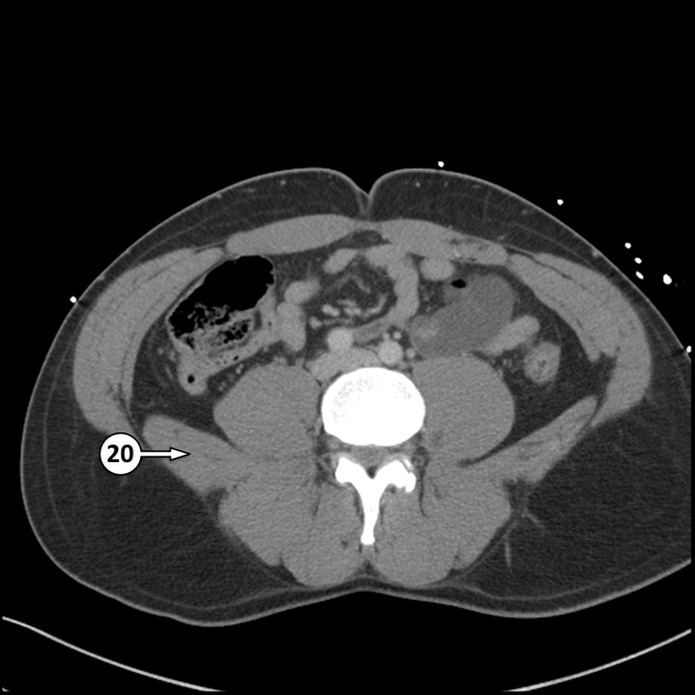
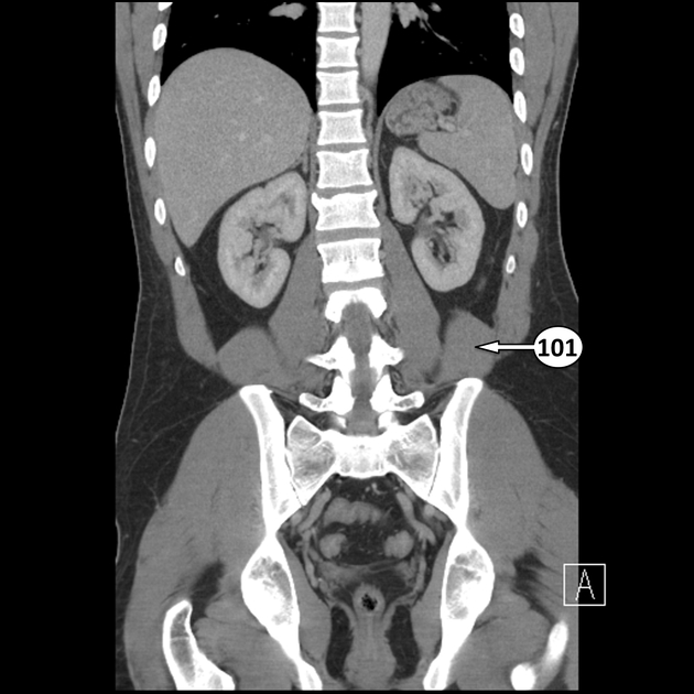
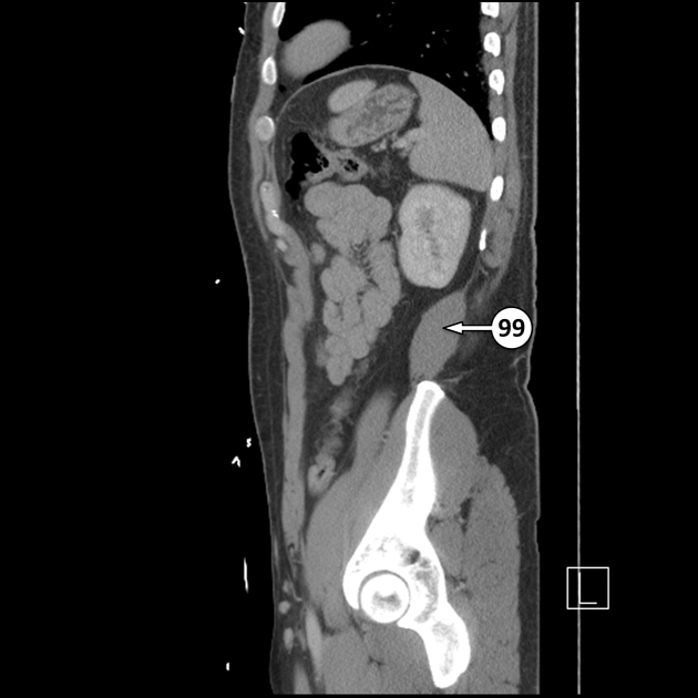
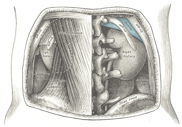
Quadratus lumborum muscle
Citation, DOI, disclosures and article data
At the time the article was created Matthew Jarvis had no recorded disclosures.
View Matthew Jarvis's current disclosuresAt the time the article was last revised Dan Mahon had no financial relationships to ineligible companies to disclose.
View Dan Mahon's current disclosures- Quadratus lumborum muscles
The quadratus lumborum muscle is a paired, irregular quadrilateral muscle that forms part of the posterior abdominal wall.
On this page:
Summary
origin: transverse process L5 vertebra, iliolumbar ligament and internal rim iliac crest
insertion: transverse processes L1-L4, inferior margin of medial half 12th rib
action: extension and lateral flexion of vertebral column; fixes 12th rib during inspiration to stabilize the diaphragm
blood supply: branches of the lumbar arteries and other smaller arterial branches, as described below
innervation: ventral rami of the 12th thoracic nerve and L1-L4 spinal nerves
Gross anatomy
Relations
The muscle is a thick, irregular, quadrilateral-shaped muscle sheet that lies in the posterior abdominal wall on each side of the lumbar vertebrae. It is superficial to the psoas major muscle.
Anterior relations include:
psoas muscle (major and minor)
Arterial supply
The quadratus lumborum muscle is supplied by:
branches of the lumbar arteries
the arteria lumbalis ima
the lumbar branch of the iliolumbar artery
branches of the subcostal artery
Innervation
ventral rami of the 12th thoracic nerve
L1-L4 spinal nerves
Action
Multiple actions, including:
-
muscle of inspiration
fixation of the last rib
-
stabilization of lower attachments of the diaphragm
proposed to provide a base for controlled diaphragmatic relaxation to facilitate precise adjustments required for speech and singing 1
when one muscle contracts, lateral flexion of vertebral column
when both muscles contract, extension of the lumbar spine
Variant anatomy
extent of attachment to the last rib varies
Related pathology
implications in unilateral lower back pain
may be enlarged in cricket fast bowlers who injure their L4 pars interarticularis 3
References
- 1. NA. Gray's anatomy. Elsevier. ISBN:0808923714. Read it at Google Books - Find it at Amazon
- 2. Moore KL, Dalley AF, Agur AM. Clinically Oriented Anatomy. Lippincott Williams & Wilkins. ISBN:1451119453. Read it at Google Books - Find it at Amazon
- 3. Engstrom CM, Walker DG, Kippers V et-al. Quadratus lumborum asymmetry and L4 pars injury in fast bowlers: a prospective MR study. Med Sci Sports Exerc. 2007;39 (6): 910-7. doi:10.1249/mss.0b013e3180408e25 - Pubmed citation
Incoming Links
Related articles: Anatomy: Spine
-
osteology
- vertebrae
- spinal canal
- cervical spine
- thoracic spine
- lumbar spine
- sacrum
- coccyx
-
anatomical variants
- vertebral body
- neural arch
- transitional vertebrae
- ossicles
- ossification centers
- intervertebral disc
- articulations
- ligaments
- musculature of the vertebral column
- muscles of the neck
- muscles of the back
-
suboccipital muscle group
- rectus capitis posterior major muscle
- rectus capitis posterior minor muscle
- obliquus capitis superior muscle
- obliquus capitis inferior muscle
- splenius capitis muscle
- splenius cervicis muscle
- erector spinae group
- transversospinalis group
- quadratus lumborum muscle
-
suboccipital muscle group
- spinal meninges and spaces
-
spinal cord
- gross anatomy
-
white matter tracts (white matter)
- corticospinal tract
- anterolateral columns
- lateral columns
-
dorsal columns
- fasiculus gracilis (column of Goll)
- fasiculus cuneatus (column of Burdach)
- grey matter
- nerve root
- central canal
- functional anatomy
- spinal cord blood supply
- sympathetic chain
Related articles: Anatomy: Abdominopelvic
- skeleton of the abdomen and pelvis
- muscles of the abdomen and pelvis
- spaces of the abdomen and pelvis
- anterior abdominal wall
- posterior abdominal wall
- abdominal cavity
- pelvic cavity
- perineum
- abdominal and pelvic viscera
- gastrointestinal tract
- spleen
- hepatobiliary system
-
endocrine system
-
adrenal gland
- adrenal vessels
- chromaffin cells
- variants
- pancreas
- organs of Zuckerkandl
-
adrenal gland
-
urinary system
-
kidney
- renal pelvis
- renal sinus
- avascular plane of Brodel
-
variants
- number
- fusion
- location
- shape
- ureter
- urinary bladder
- urethra
- embryology
-
kidney
- male reproductive system
-
female reproductive system
- vulva
- vagina
- uterus
- adnexa
- Fallopian tubes
- ovaries
- broad ligament (mnemonic)
- variant anatomy
- embryology
- blood supply of the abdomen and pelvis
- arteries
-
abdominal aorta
- inferior phrenic artery
- celiac artery
- superior mesenteric artery
- middle suprarenal artery
- renal artery (variant anatomy)
- gonadal artery (ovarian artery | testicular artery)
- inferior mesenteric artery
- lumbar arteries
- median sacral artery
-
common iliac artery
- external iliac artery
-
internal iliac artery (mnemonic)
- anterior division
- umbilical artery
- superior vesical artery
- obturator artery
- vaginal artery
- inferior vesical artery
- uterine artery
- middle rectal artery
-
internal pudendal artery
- inferior rectal artery
-
perineal artery
- posterior scrotal artery
- transverse perineal artery
- artery to the bulb
- deep artery of the penis/clitoris
- dorsal artery of the penis/clitoris
- inferior gluteal artery
- posterior division (mnemonic)
- variant anatomy
- anterior division
-
abdominal aorta
- portal venous system
- veins
- anastomoses
- arterioarterial anastomoses
- portal-systemic venous collateral pathways
- watershed areas
- arteries
- lymphatics
- innervation of the abdomen and pelvis
- thoracic splanchnic nerves
- lumbar plexus
-
sacral plexus
- lumbosacral trunk
- sciatic nerve
- superior gluteal nerve
- inferior gluteal nerve
- nerve to piriformis
- perforating cutaneous nerve
- posterior femoral cutaneous nerve
- parasympathetic pelvic splanchnic nerves
- pudendal nerve
- nerve to quadratus femoris and inferior gemellus muscles
- nerve to internal obturator and superior gemellus muscles
- autonomic ganglia and plexuses


 Unable to process the form. Check for errors and try again.