
The axis is the second cervical vertebra, commonly called C2. It is an atypical cervical vertebra with unique features and important relations that make it easily recognisable. Its most prominent feature is the odontoid process (also know as the dens or peg), which is embryologically the body of the atlas (C1) 1,2. It plays an important role in rotation of the head with the majority of movement occurring around the dens and at the atlanto-axial joint. There are five primary and two secondary ossification centers which are discussed below in more detail 2-4.
On this page:
Gross anatomy
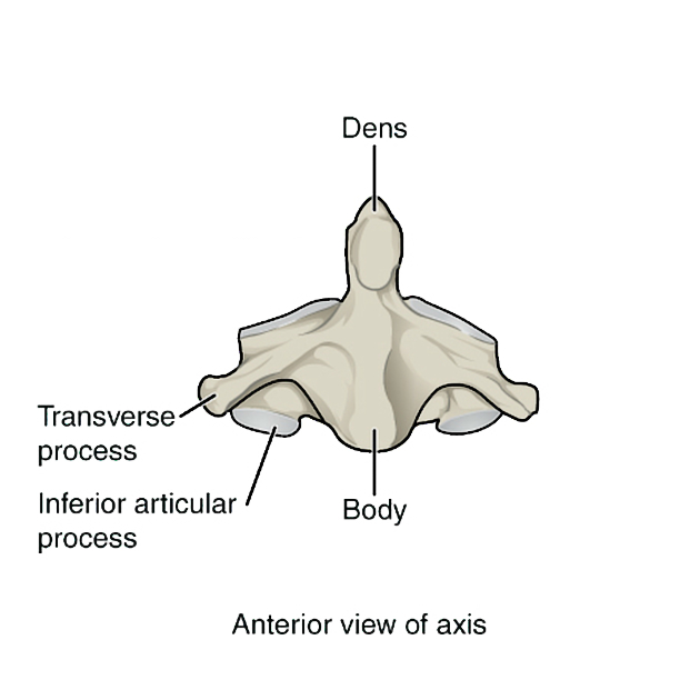
The axis is formed by a body with the attached dens, two lateral masses, a posterior vertebral arch (formed by the pedicle and a thick lamina), and a large spinous process, which is commonly bifid.
Anterior components of the axis are composed of:
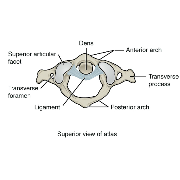
odontoid process (dens or peg): conical in shape, projects up from the body, smooth posterior surface
-
body
in skeletally mature individuals, there is almost always a lucent or sclerotic remnant of the synchondrosis between the body and dens
lateral mass bears the weight of skull and transfers through to C3 vertebral body
transverse process with foramina transversarium, L-shaped, directed up and out to allow lateral bend in vertebral artery
superior articular facets, slopes down from body like shoulders, extends over pedicles and lateral masses
inferior articular facets, face anteroinferiorly like typical cervical vertebra
Posterior elements of the axis are composed of:
pedicle
lamina, thick and rounded
spinous process, large and bifid
Articulations
-
atlantoaxial joint
-
median:
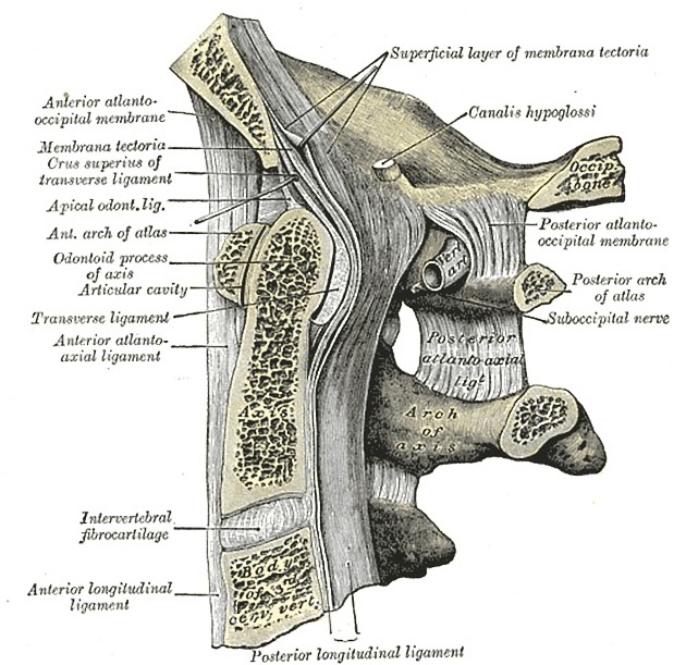
synovial with joint capsule and bursa between posterior dens and cruciform ligament
articulation: anterior surface of dens with the posterior surface of anterior arch of C1, surfaces covered in hyaline cartilage
movement: head rotation, flexion/extension
-
lateral
synovial with joint capsule
articulation: inferior articular facet of C1 with superior articular facet of C2, surfaces covered in hyaline cartilage
movement: head rotation, flexion/extension
-
C2/3 uncovertebral joint
inferior articular facets of C2 with superior articular facets of C3 (facet joints)
intervertebral joint with C3 via the C2/C3 intervertebral disc
Attachments
-
musculotendinous
-
attached to the anterior surface of the vertebral body
longus colli
-
attached to transverse processes
levator scapulae
scalenus medius
splenius cervicis
-
attached to spinous processes
semispinalis cervicis
rectus capitis posterior major
obliquus capitis inferior
-
attached to posterior surface of lamina
multifidus and longissimus
-
-
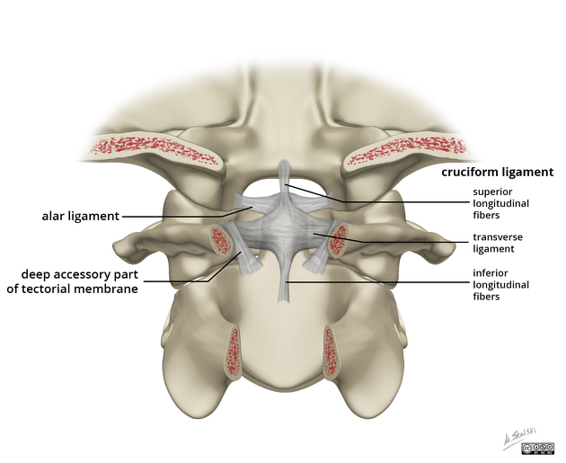
tectorial membrane
extends upward in continuity with posterior longitudinal ligament
attached to back of body of C2 and anterior margin of foramen magnum
-
ligaments
-
apical ligament, embryological remnant of the notochord
apex of dens to anterior margin of foramen magnum
-
paired alar ligaments
sloping upper margin of dens to margin of foramen magnum
-
cruciform ligament with synovial bursa and fibrous capsule
longitudinal part - attached from the body of the axis to the basiocciput
transverse part - laterally attached to the medial aspect of the lateral masses of C1, provides the most stability retaining the dens in contact with the atlas (C1)
found between the tectorial membrane and apical ligament
accessory atlantoaxial ligament: from posterior body of C2 to lateral mass of C1
anterior atlantoaxial membrane: from the body of the C2 to the anterior arch of C1
posterior atlantoaxial membrane (upper part of the ligamentun flavum between C1 and C2)
ligamentum nuchae
anterior longitudinal ligament: passes anterior to the anterior atlantoaxial membrane before attaching to the anterior tubercle of C1
-
Relations
Anteriorly
anterior atlantoaxial membrane
Posteriorly
posterior atlantoaxial membrane
-
posterior to dens
-
important ligamentous structures are attached to the dens and play an important role in C1/C2 stability
apical ligament: embryological remnant of the notochord
accessory ligament
tectorial membrane
-
Laterally
-
vertebral arteries and veins
traverse foramina transversarium moving laterally
Supero-posteriorly
Centrally
spinal cord traverses vertebral foramen
basivertebral veins
Variant anatomy
hypoplasia
aplasia (rare)
bifid spinous process (common)
bicornuate odontoid
basilar invagination - upward displacement of the tip of odontoid process above foramen magnum
See also: vertebral anomalies.
Radiographic features
Plain radiograph
the odontoid process and atlanto-axial joint are best appreciated in an AP open mouth view
soft tissue contours are visible on lateral views
Development
There are five primary and two secondary ossifications centers in the axis (C2).
Primary ossification centers:
-
vertebral arches
two primary centers
appear at 7-8 weeks
fuses with the odontoid and centrum at 3 to 6 years 8
-
vertebral centrum/body
one primary center
appear at 4-5 months
-
dens (odontoid process)
bilateral centers
appears around 6 months
both centers fuse before birth forming conical mass
fuse with the centrum by 3 to 6 years at the rim of subdental synchrondosis 8,9 . In some people, the rim may not completely ossify until 12 years of age. The central portion of the synchrondosis may not ossify throghout the lifetime of a patient 9
Secondary ossification centers:
-
apex of dens / os terminale
from cuneiform cartilage
-
variable appearance and fusion
most commonly appear from 3 to 6 years of age 8,9
fuse at around the 12th year 9
-
dens separated from the vertebral body by a cartilaginous disc
circumference ossifies 3,5


 Unable to process the form. Check for errors and try again.