
Pars interarticularis
Citation, DOI, disclosures and article data
At the time the article was created Craig Hacking had no recorded disclosures.
View Craig Hacking's current disclosuresAt the time the article was last revised Tariq Walizai had no financial relationships to ineligible companies to disclose.
View Tariq Walizai's current disclosures- pars
The pars interarticularis or simply pars (plural: partes interarticulares) is the part of a vertebra located between the superior and inferior articular processes.
On this page:
Gross anatomy
In the axis and the thoracolumbar spine, the pars interarticularis is a narrow isthmus between the superior and inferior facets. In the subaxial cervical spine, the pars interarticularis equivalent is termed the lateral mass or articular pillar.
In the axis, the pars interarticularis is located anterolateral to the spinal canal 5. In the thoracolumbar spine, the pars interarticularis is located posterolateral to the spinal canal.
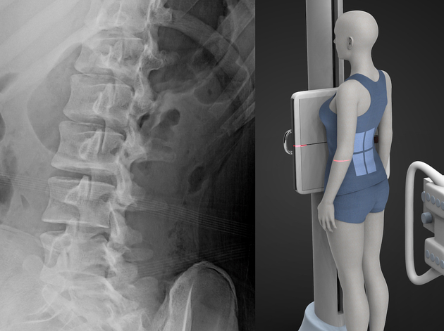
Radiographic features
In the axial plane, the pars is located at the junction of the pedicle and lamina.
In the oblique lumbar radiograph, the neck of the Scottie dog represents the pars.
Related pathology
pars defect (spondylolysis)
Quiz questions
References
- 1. Moore KL, Agur AMR, Dalley AF. Clinically oriented anatomy. LWW. ISBN:1451119453. Read it at Google Books - Find it at Amazon
- 2. Last's anatomy, regional and applied. Churchill Livingstone. ISBN:044304662X. Read it at Google Books - Find it at Amazon
- 3. Butler P, Mitchell A, Healy JC. Applied Radiological Anatomy. Cambridge University Press. (2012) ISBN:0521766664. Read it at Google Books - Find it at Amazon
- 4. Susan Standring. Gray's Anatomy. ISBN: 9780702052309
- 5. Ebraheim N, Fow J, Xu R, Yeasting R. The Location of the Pedicle and Pars Interarticularis in the Axis. Spine (Phila Pa 1976). 2001;26(4):E34-7. doi:10.1097/00007632-200102150-00002 - Pubmed
Incoming Links
- Pars interarticulares stress reaction (SPECT-CT bone scan)
- Bilateral L4 spondylolysis
- Grade II anterolisthesis of L5 over S1
- Oppenheimer ossicle
- Chalk stick fracture
- Oppenheimer ossicle
- Collar sign in spondylolysis
- Pars interarticularis defect
- Pars interarticularis defect
- Pars interarticularis defect
- Pars interarticularis defect
- Spondylolisthesis, spondylolysis and spondylosis, with osteoporosis and bilateral L5 sacralisation
- Pars interarticularis fracture
- Pars interarticularis
- Bilateral pars defect
Related articles: Anatomy: Spine
-
osteology
-
vertebrae
- vertebral vascular foramen
- pars interarticularis
- intervertebral foramen
- spinal canal
- cervical spine[+][+]
- thoracic spine[+][+]
- lumbar spine[+][+]
- sacrum
- coccyx
-
anatomical variants[+][+]
- vertebral body
- neural arch
- transitional vertebrae
- ossicles
- ossification centers
-
vertebrae
- intervertebral disc[+][+]
- articulations[+][+]
- ligaments[+][+]
- musculature of the vertebral column[+][+]
- muscles of the neck
- muscles of the back
-
suboccipital muscle group
- rectus capitis posterior major muscle
- rectus capitis posterior minor muscle
- obliquus capitis superior muscle
- obliquus capitis inferior muscle
- splenius capitis muscle
- splenius cervicis muscle
- erector spinae group
- transversospinalis group
- quadratus lumborum muscle
-
suboccipital muscle group
- spinal meninges and spaces[+][+]
-
spinal cord[+][+]
- gross anatomy
-
white matter tracts (white matter)
- corticospinal tract
- anterolateral columns
- lateral columns
-
dorsal columns
- fasiculus gracilis (column of Goll)
- fasiculus cuneatus (column of Burdach)
- grey matter
- nerve root
- central canal
- functional anatomy
- spinal cord blood supply
- sympathetic chain[+][+]


 Unable to process the form. Check for errors and try again.