
Wall-echo-shadow sign (ultrasound)
Citation, DOI, disclosures and article data
Citation:
Shin J, Murphy A, Jones J, et al. Wall-echo-shadow sign (ultrasound). Reference article, Radiopaedia.org (Accessed on 09 Mar 2025) https://doi.org/10.53347/rID-21822
Permalink:
rID:
21822
Article created:
20 Feb 2013,
James Shin
Disclosures:
At the time the article was created James Shin had no recorded disclosures.
View James Shin's current disclosures
Last revised:
Disclosures:
At the time the article was last revised Andrew Murphy had no financial relationships to ineligible companies to disclose.
View Andrew Murphy's current disclosures
Revisions:
14 times, by
11 contributors -
see full revision history and disclosures
Systems:
Sections:
Tags:
Synonyms:
- WES sign
- Wall echo shadow sign
- Double-arc-shadow sign
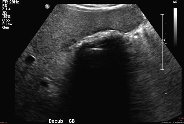
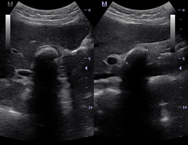
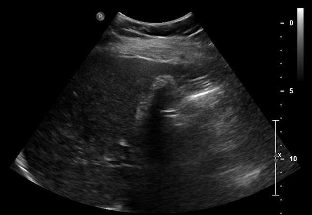
The wall-echo-shadow sign (also known as WES sign) is an ultrasonographic finding within the gallbladder fossa referring to the appearance of a "wall-echo-shadow":
- a curvilinear hyperechogenic line representing the gallbladder wall
- a thin hypoechoic space representing a small amount of bile
- a curvilinear hyperechogenic line representing the near surface of gallstone(s)
- and acoustic shadowing distal to the surface of the gallstone(s)
The sign suggests either a large gallstone or multiple small gallstones filling the lumen of a contracted or incompletely visualized gallbladder. Recognizing this finding helps to avoid misinterpretation of a stone-filled gallbladder as a loop of bowel 3.
Differential diagnosis
- air-filled loop of bowel
- porcelain gallbladder: lacks the thin hypoechoic bile space between the wall and gallstone, as seen in the wall-echo-shadow sign
- emphysematous cholecystitis
References
- 1. Rybicki F. The WES Sign. Radiology. 2000;214(3):881-2. doi:10.1148/radiology.214.3.r00mr38881
- 2. MacDonald F, Cooperberg P, Cohen M. The WES Triad — A Specific Sonographic Sign of Gallstones in the Contracted Gallbladder. Gastrointest Radiol. 1981;6(1):39-41. doi:10.1007/bf01890219
- 3. William E. Brant, Clyde A. Helms. The Brant and Helms Solution: Fundamentals of Diagnostic Radiology, Third Edition, Plus Integrated Content Website (4 Vol. Set). (2006) ISBN: 0781765188
Incoming Links
Multiple choice questions:
Related articles: Ultrasound
-
ultrasound
- ultrasound signs
- physics and imaging modes
- grey-scale (B-mode)
- motion mode (M-mode)
- color flow Doppler (CFD)
-
spectral Doppler
- pulsed wave Doppler (PWD)
- continuous wave Doppler (CWD)
- superb microvascular imaging (SMI)
- tissue Doppler imaging (TDI)
- obstetric ultrasound
- fetal morphology assessment
- gynecologic ultrasound
- vascular ultrasound
- carotids
- extremities
- breast ultrasound
-
musculoskeletal ultrasound
- technique/artifacts
- ultrasound of arthropathies
- other
- pediatric musculoskeletal ultrasound
- ankle/foot ultrasound
- knee ultrasound
- hip ultrasound
- hand ultrasound
- wrist ultrasound
- elbow ultrasound
- shoulder ultrasound
- liver ultrasound
- gallbladder ultrasound
- pancreatic ultrasound
- gastrointestinal ultrasound
- testicular and scrotal ultrasound
- prostate ultrasound
- transrectal ultrasound (TRUS)
- neck and thyroid ultrasound
- echocardiography
- speckle tracking echocardiography
- fetal echocardiography
- contrast-enhanced echocardiography
- epicardial echocardiography
- three dimensional (3D) echocardiography
- transesophageal echocardiography (TEE)
- transthoracic echocardiography (TTE)
- left ventricular systolic and diastolic function
- structure and morphology
- systolic function
- diastolic function
- right ventricular assessment
- right and left atria
- hemodynamics
- ultrasound interventions
- ultrasound-guided biopsy
- core biopsy
- ultrasound-guided percutaneous drainage
- ultrasound-guided musculoskeletal interventions
- joint injection
- nerve blocks
- ultrasound-guided intravenous cannulation


 Unable to process the form. Check for errors and try again.