Radiopaedia Blog

The introduction of improved playlists earlier this year has been a superb advancement in functionality and user experience. Be it for organising ones own cases, sharing with friends and colleagues, or teaching at institutional or national meetings.

At present playlists are neither searchable or indexed, but contributors will often notice that over time some of their cases have been included in other users' playlists. When viewing a case which is included in a playlist, you can navigate to that playlist. From there you may explore new cases and from them additional playlists, opening up a kaleidoscope of educational merriment.

It provides a proud individual moment, as well as a sense of satisfaction, when others are not only appreciating, but also in many cases using your cases to educate others across the globe.

Below are a selection of some of the awesome playlists that are emerging.

Playlists from our courses

Chest module from our Trauma course

This is one of the playlists that was used for the recent Radiopaedia.org trauma course by managing editor Andrew Dixon. view here

Each case is attributed clearly, so that everyone can see your contribution. Even better, all contributors whose cases are included in these courses are given complimentary access to the whole online course.

ANZSNR Neuroradiology series

Alternatively it may feature in national meeting presentations like the ANZSNR Neuroradiology series curated by Radiopaedia.org founder Frank Galliard. view here

CT brain for Emergency Physicians

Playlists can be very personal or very public. They may be long or succinct. The topic may be broad and targeted at the clinician community like this tidy playlist on CT Head for Emergency Physicians. view here

Collaborative projects

On the other hand they may be immensely focussed, such as the list created to aid the radiopaedia.org team in the production of a collaborative poster for the 2014 RANZCR meeting entitled, "Wrong place, wrong time: Imaging and embryological features of congenital renal anomalies". view here

Personal collections

Playlists can also be dynamic, changing on a day to day basis, like some of my own collections for individual departments in my own little hospital (RIPAS) in Brunei. view here

Exam preparation

Finally in keeping with the spirit of both participation and educational contribution a plethora of playlists are out there to help others in exam preparation. This includes some superb sets from managing editor Jeremy Jones, like this pot pouri of paediatric MSK cases for the UK Royal College of Radiologists FRCR 2B examination. view here

Explore, enjoy, share and educate.

Now that we have over 20,000 cases, it has become imperative that we are able to more easily find quality cases with confirmed diagnoses. To that end over the next week or two we are introducing a new component to all cases: Diagnostic Certainty (DC). 

This new user interface is located immediately below the title in case edit mode. 

There are 5 possible levels of diagnostic certainty 

  1. Possible
    • The preferred diagnosis is one of a number of possible diagnoses, and although it is perhaps more likely, other conditions remain viable alternatives. Cases like this are sometimes worthwhile, but often they are not. What you need to do: include a discussion on why you favour your diagnosis. Be prepared for your case to be rejected for publication. That's ok, you can always keep it as one of your unlisted cases.
  2. Probable
    • The preferred diagnosis is far more likely (>90%) than any alternative, but the diagnosis has not been established.
  3. Confirmed but unsubstantiated
    • You have reason to believe the diagnosis was confirmed, but you do not have access to the proof and cannot upload it. For example, you collected this case some time ago and have included "path proven" in the description but no actual histology report. Alternatively the referral states the diagnosis which was established elsewhere.
  4. Confirmed and substantiated
    • The diagnosis is known beyond a doubt and proof is included in this case. For example, histology report +/- slides are included, or confirmatory tests have been provided and those results included. Sometimes images are pathognomonic (e.g. fractures) and need no supportive evidence.
  5. Not applicable
    • The images are not of a patient with a particular diagnosis. For example, they may be a normal scan for teaching purposes, or alternatively a diagram or flow chart.

How diagnostic certainty is used on the site

The diagnostic certainty of a case has many implications and functions. 

Firstly it has a strong influence on case completeness, and therefore sorting search results by case completeness will favour cases with high DC. Similarly these cases will be more frequently shown in Quiz Mode. 

Secondly search results will show the DC state as a coloured dot (matching the slider). 

Thirdly search results can be (soon) filtered to only show cases of a minimum DC (e.g. at least confirmed but unsubstantiated). 


Have you registered for our Adult Brain MRI Review Course yet? You can watch all six hours of live recorded content for just $50 or for free in 115 developing countries. Don't miss your chance to watch this special event. 

The Diagnostic Imaging Pathways App is an offline decision-support tool designed to help clinicians make appropriate medical imaging choices. This free resource is based upon the popular website of the same name. It's currently available for iOS, with an android version expected soon.

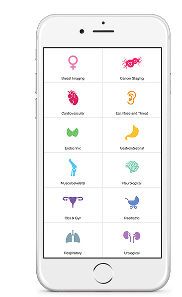
The App is divided into 12 subspecialty categories under which specific clinical pathways can be accessed to guide the user towards the most appropriate imaging test. An overview button allows each pathway to be visualised in its entirety at any time.

There are many things to like about this App, particularly the fact that it is a free resource made available globally. Trying to cover every single clinical scenario is however an impossible task and keeping pathways up to date and evidence based is going to prove a constant challenge. 

In order to get an idea of the current status of this App and its potential for the future, we invited a group of radiologists on Twitter to take a look for us. We'd love to get your feedback too, which you can send to us through the comments section or via social media. 

@DrAndrewDixon


Erik Ranschaert

Review: 
This mobile application is a very useful tool which every hospital-based physician should have at hand in order to help them choose the most appropriate diagnostic approach. The form is very attractive but some of the content runs a bit behind, which is a little disappointing. The information in some pathways would benefit from more detailed and updated information.

The prostate pathway in particular seems very old fashioned. The initial recommended imaging modality is TRUS biopsy although many cancers are being missed using this technique. MRI is really of great added value in case of negative TRUS biopsies as tumours in the transition zone are easily missed, and this is not reflected in the pathway. And who is still performing endorectal MRI? The most appropriate pathway is normal (transabdominal) prostate MRI (preferably 3T).

Rating: 


Jeremy Jones

Review:
The app covers a broad selection of presentations across a number of paediatric specialties. The topics covered are representative of the majority of the presentations I see on a daily basis and the suggestions for imaging are relevant for the UK as well as Australia & New Zealand. The ability to see an overview of the guidance as well as having a pathway that requires interaction is helpful to gain an understanding of the whole pathway as well as the arm that you are using. Overall, I think that this will be a helpful resource for any healthcare professional who uses radiological services.

Rating:


Jenny Hoang

Review:
A good start, but there’s a lot more work to do! The app was easy to use and graphics were cute. Overview of pathways was great to have. I do worry that this app could be perceived as evidence-based guidelines when there are actually major deficiencies. I found many of the head and neck pathways to be incomplete, too simplified or out of date. 

I particularly disagree with the incidental thyroid nodule pathway which recommends either US or thyroid scintigraphy for all nodules depending on TSH. This approach is not cost-effective and can lead to harm from over-investigation and overdiagnosis. The approach in the ACR White Paper on Incidental Thyroid Findings should be used instead. There is also no mention of the use of 4D-CT in the hyperparathyroidism pathway and the radiation dose from parathyroid scintigraphy is much more than the 1-5mSv listed. 

Rating:


Vikas Shah

Review:
The Diagnostic Imaging Pathways (DIP) app is visually pleasing and easy to navigate, with the pathway overview in particular being a handy feature. Some key guidelines are missing (e.g. anal cancer staging) and some take a path quite different to that in most UK centres (e.g. recommending both abdominal AXR and CT if acute diverticulitis is suspected, or contrast enhanced CT in renal colic if the patient is aged over 50). However, the concept is sound, and the app is technically well designed so I can see this being "spun off" in different countries to suit local needs. 

Rating:


Nikhil Mehta

Review:
The diagnostic imaging pathways app has a very user friendly, easy and fast on the go interface. I tried various clinical scenarios, which I come across in day to day radiology practice. Some pathways work along the same line as we use India. Some of the pathways need updating, like the criteria for multiple sclerosis. The stroke pathway matches our evaluation steps. We use the ASPECTS score for MCA infarction and CT perfusion most often in the proper clinical scenario followed by extracranial carotid angiography. Overall I think the pathways are useful but further additions and updating are required.

Rating:


Alexandra Stanislavski

Review:
The DIP website and smartphone app are designed assist clinicians with a) choosing the best imaging tests for their patient, and b) adjusting patient management depending of the test result.  Among the sixteen pathways in the O&G section, there are some that achieve this very well. For example,  “Adnexal masses” and “Bleeding in early pregnancy” are excellent, detailed and current pathways. Several others however are less satisfying because they’re either too brief, or do not clearly define the role of imaging.  Interestingly, the website flags many of these as “due for review October 2015”. I am looking forward to seeing the updates.
 
The transition from website to app is a great idea, and the app menu interface is nicely presented. I would have liked to see more focus on the “Pathway Overview”, rather than multi-page decision making tool, which is a little clumsy. The best bits on the website are the pearls of wisdom in the hover boxes on the pathway overview page, and these are lost in the app version. Overall – great concepts, impressive breadth – with room for improvement for content and app usability.

Rating:


20th Jul 2015 23:01 UTC

New features - July 2015

This week we have released a bunch of small improvements to the site, some of which represent significant improvements to the back end of the site. 

Case view

The largest release is that we have replaced the case viewing engine to one similar to that used in presentation mode. This now means:

  1. stacks will start loading from the key image (or middle image if a key image has not been selected); this means that you will get to the important part of the stack straight away rather than having to start at the very top (often blank) image. 
  2. scrolling will be touch responsive on mobile devices (phones / tablets); this will be a huge improvement as previously scrolling was impractical. 

rID

We have instituted a unique radiopaedia ID (rID) for all cases and exposed this in our case information box (to the right of the case images). 

The box is now collapsed by default, as we plan to put all sorts of additional info in there which is not needed for general browsing. Keeps things nice and neat. 

The rID is an excellent way to keep track of where you got images from in a presentation. 

To make this even easier we have also included rID in the 'download image' attribution links. 


Presentation mode

Presentation mode has had a couple of minor tweaks. Firstly the size of the text has been increased (1) and will scale according to your screen size. This will be easier on the eye and much better when presenting cases on projectors. 

Secondly we have changed the attribution at the bottom right corner. The contributing user's name (2) is now a blue clickable link to the contributor's profile, their supporter status is indicated (3) and the rID is exposed (4). Clicking the rID takes you to the case in view mode. 

There is much more to come, so stay tuned for an every bigger and better radiopaedia.org

Thanks for your continued support. 

Frank. 

Blog Subscription

We will only send you an email when there are new posts.

Updating… Please wait.

 Unable to process the form. Check for errors and try again.

 Thank you for updating your details.