
CPPD of the cervical spine
Updates to Case Attributes
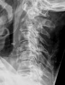
Calcium pyrophosphate dihydrate (CPPD) disease or pseudogout of the cervical spine.
A partially calcified "mass" is seen behind the odontoid process compressing the cervical cord, so-called "crowned dens syndrome".
It is an often misrecognized cause of acute neck pain in the elderly. The pseudotumour behind the dens may cause cervicomedullary compression.
Typical radiographic manifestations of CPPD in the cervical spine:
Calcific deposits can be seen in:
- the annulus fibrosus and
transversetransverse ligament of the dens - the supraspinous and interspinous ligaments
- the intervertebral discs
- the longitudinal ligaments (may cause spinal stenosis)
- capsular calcifications in the apophyseal facet joints
- pseudotumorous peri-odontoid mass of the foramen magnum ("crowned dens")
Pyrophosphate arthropathy (in long-standing disease) causing:
- narrowing of intervertebral discs
- bone sclerosis
- osteophyte formation
- depositions of crystals within the joints can lead to erosions, subchondral cysts and pathologic fractures
- erosions and cysts of the odontoid process
The differential for calcific deposits in the cervical spine is a limited one:
- Hydroxyapatite deposition disease (HADD) - can be indistinguishable from CPPD on CT
- The inflammatory response to the crystal deposits in the disc space can be aggressive enough to mimic a discitis
- Gout can cause similar findings
Clinical history and/or crystal analysis may be needed for the definitive diagnosis. In difficult cases a biopsy may be needed.
-<p><strong><a href="/articles/cppd" title="CPPD">Calcium pyrophosphate dihydrate (CPPD) disease</a></strong> or <a href="/articles/cppd" title="Pseudogout"><strong>pseudogout</strong></a> of the cervical spine.</p><p>A partially calcified "mass" is seen behind the odontoid process compressing the cervical cord, so-called "<a href="/articles/crowned-dens-syndrome" title="crowned dens syndrome">crowned dens syndrome</a>". </p><p>It is an often misrecognized cause of acute neck pain in the elderly. The pseudotumour behind the dens may cause cervicomedullary compression.</p><p>Typical radiographic manifestations of CPPD in the cervical spine:</p><p><em>Calcific deposits can be seen in:</em></p><ul><li>the annulus fibrosus and transverse ligament of the dens</li><li>the supraspinous and interspinous ligaments</li><li>the intervertebral discs</li><li>the longitudinal ligaments (may cause spinal stenosis)</li><li>capsular calcifications in the apophyseal facet joints</li><li>pseudotumorous peri-odontoid mass of the foramen magnum ("crowned dens")</li></ul><em>Pyrophosphate arthropathy (in long-standing disease) causing:</em><ul><li>narrowing of intervertebral discs</li><li>bone sclerosis</li><li>osteophyte formation</li><li>depositions of crystals within the joints can lead to erosions, subchondral cysts and pathologic fractures</li><li>erosions and cysts of the odontoid process</li></ul><p>The differential for calcific deposits in the cervical spine is a limited one:</p><ul><li><strong><a href="/articles/hadd" title="HADD">Hydroxyapatite deposition disease (HADD)</a></strong> - can be indistinguishable from CPPD on CT</li><li>The inflammatory response to the crystal deposits in the disc space can be aggressive enough to mimic a <strong><a href="/articles/spondylodiscitis" title="Spondylodiscitis">discitis</a></strong></li><li><strong><a href="/articles/gout" title="Gout">Gout</a> </strong>can cause similar findings</li></ul><p>Clinical history and/or crystal analysis may be needed for the definitive diagnosis. In difficult cases a biopsy may be needed.</p>- +<p><strong><a href="/articles/cppd">Calcium pyrophosphate dihydrate (CPPD) disease</a></strong> or <a href="/articles/cppd"><strong>pseudogout</strong></a> of the cervical spine.</p><p>A partially calcified "mass" is seen behind the odontoid process compressing the cervical cord, so-called "<a href="/articles/crowned-dens-syndrome">crowned dens syndrome</a>". </p><p>It is an often misrecognized cause of acute neck pain in the elderly. The pseudotumour behind the dens may cause cervicomedullary compression.</p><p>Typical radiographic manifestations of CPPD in the cervical spine:</p><p><em>Calcific deposits can be seen in:</em></p><ul>
- +<li>the annulus fibrosus and transverse ligament of the dens</li>
- +<li>the supraspinous and interspinous ligaments</li>
- +<li>the intervertebral discs</li>
- +<li>the longitudinal ligaments (may cause spinal stenosis)</li>
- +<li>capsular calcifications in the apophyseal facet joints</li>
- +<li>pseudotumorous peri-odontoid mass of the foramen magnum ("crowned dens")</li>
- +</ul><p><em>Pyrophosphate arthropathy (in long-standing disease) causing:</em></p><ul>
- +<li>narrowing of intervertebral discs</li>
- +<li>bone sclerosis</li>
- +<li>osteophyte formation</li>
- +<li>depositions of crystals within the joints can lead to erosions, subchondral cysts and pathologic fractures</li>
- +<li>erosions and cysts of the odontoid process</li>
- +</ul><p>The differential for calcific deposits in the cervical spine is a limited one:</p><ul>
- +<li>
- +<strong><a href="/articles/hadd">Hydroxyapatite deposition disease (HADD)</a></strong> - can be indistinguishable from CPPD on CT</li>
- +<li>The inflammatory response to the crystal deposits in the disc space can be aggressive enough to mimic a <strong><a href="/articles/spondylodiscitis">discitis</a></strong>
- +</li>
- +<li>
- +<strong><a href="/articles/gout">Gout</a> </strong>can cause similar findings</li>
- +</ul><p>Clinical history and/or crystal analysis may be needed for the definitive diagnosis. In difficult cases a biopsy may be needed.</p>
Updates to Study Attributes
Image X-ray (Lateral) ( update )
Updates to Study Attributes
CT cervical spine coronal view: erosive changes at the apophyseal joints of C1-C2 with subluxation of the atlanto-axialatlantoaxial joint, discdisc narrowing and erosive changes at the C2-3 level, extensive disc space narrowing at all levels. Narrowing and erosive changes at the facet joints. Calcified mass behind the dens.
CT cervical spine lateral view: partially calcified pseudomasspseudo mass behind the dens, extensive narrowing and erosive changes of the end-plates at the C2-C3 level, largemassive erosion anteriorly in the C3 body. Intervertebral disc calcifications at the C6-C7 and C7-Th1 levels, calcifications in the posterior and anterior longitudinal ligaments and annulus fibrosus, calcifications at the interspinous ligament C7, spondylophyte formation anteriorly.
Image CT (bone window) ( update )

Image CT (bone window) ( update )

Image CT (bone window) ( update )


 Unable to process the form. Check for errors and try again.