


121,460 results
Case
Cellulitis - perineum

Published
07 Jun 2020
66% complete
Ultrasound
Article
Ciliopathies
Ciliopathies refer to diseases due to malfunctioning cilia (singular: cilium). Cilia are organelles that are external extensions of the cell membrane. Cilia fall into two main types: primary (or immotile) cilia and motile cilia.
Clinical presentation
Primary cilia are found in virtually every...
Case
Cytotoxic lesions of the corpus callosum (CLOCCs)

Published
10 Sep 2020
74% complete
MRI
Case
Popliteal deep venous thrombosis

Published
18 Apr 2011
68% complete
CT
Article
Glenoid labral tear
Glenoid labral tears are the injuries of the glenoid labrum and a possible cause of shoulder pain.
Clinical presentation
Patients with labral tears may present with a wide range of symptoms (depends on the injury type), which are often non-specific:
pain or discomfort (usually a precise point...
Case
Acute colonic diverticulitis

Published
05 Nov 2023
73% complete
CT
Article
Shoulder (supine lateral scapula view)
The supine lateral scapula view (anterior oblique AP) is a modified lateral shoulder projection often utilised in trauma imaging. Orthogonal to the AP shoulder (note so is an axillary view); It is a pertinent projection to assess suspected dislocations, scapula fractures and degenerative changes...
Article
Periosteal chondrosarcoma
Periosteal chondrosarcomas, previously also known as juxta-cortical chondrosarcomas, are cartilagineous or chondroid matrix-generating neoplasms originating in close association with the periosteum from the bony surface 1-3.
Terminology
The term ‘juxta-cortical chondrosarcoma’ is no longer rec...
Article
Subdural hygroma
Subdural hygromas (alternative plural: hygromata 9) refer to the accumulation of fluid in the subdural space. In many cases, it is considered an epiphenomenon of head injury when it is called a traumatic subdural hygroma.
Epidemiology
Subdural hygromas are encountered in all age-groups but ar...
Case
Left atrial thrombus

Published
18 Apr 2011
80% complete
CT
Case
Calcified convexity meningioma

Published
06 May 2013
74% complete
MRI
Article
Facial nerve
The facial nerve is the seventh (CN VII) cranial nerve and comprises two roots, a motor root and a smaller mixed sensory, taste and parasympathetic root, known as nervus intermedius, which join together within the temporal bone (TA: nervus facialis or nervus cranialis VII).
The facial nerve has...
Case
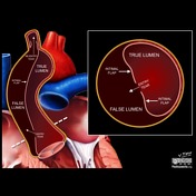
Pathogenesis of aortic dissection (illustration)
Published
08 Oct 2016
44% complete
Diagram
Playlist
1a


8 cases
No description provided
Article
Osteonecrosis of the femoral head
Osteonecrosis of the femoral head, previously known as avascular necrosis (AVN) of the hip, is the most common site for osteonecrosis, presumably due to a combination of precarious blood supply and high loading when standing.
Idiopathic osteonecrosis of the femoral head epiphysis in children (...
Article
Meningocele
Meningoceles (also spelt meningocoele) are protrusions of the meninges through a defect or weak point in the skull or spine, usually involving the soft tissues beneath the surface of the skin. They are typically categorised into congenital, iatrogenic (e.g. following a craniotomy, sinus surgery,...
Case
Ulnar fracture and radial head dislocation

Published
16 May 2016
94% complete
X-ray
Case
Foreign body in heel

Published
28 Oct 2012
66% complete
Ultrasound
Case
Cricoid cartilage fracture

Published
06 Oct 2022
92% complete
CT
Playlist
Key conditions - chest


7 cases
No description provided


 Unable to process the form. Check for errors and try again.