


121,077 results
Article
Osteolipoma
Osteolipomas, also known as ossified lipomas, are rare intracranial masses, typically located in the suprasellar cistern, composed of mature adipocytes surrounded by calcified ossification 1. They are a variant of intracranial lipomas which rarely have calcification/ossification elsewhere, with ...
Case
Glioblastoma with primitive neuronal components

Published
07 Aug 2022
95% complete
CT
Case
Arachnoid cyst - cerebellopontine angle

Published
07 Aug 2022
74% complete
MRI
Case
Left upper lobe collapse

Published
24 Oct 2020
75% complete
CT
Case
Sturge-Weber syndrome

Published
20 Oct 2010
52% complete
CT
MRI
Case
Branch retinal artery occlusion

Published
28 Apr 2022
79% complete
Photo
Article
Langerhans cell histiocytosis
Langerhans cell histiocytosis (LCH) is a rare multisystem disease with a wide and heterogeneous clinical spectrum and variable extent of involvement.
Terminology
Langerhans cell histiocytosis was previously known as histiocytosis X. The newer term is preferred as it is more descriptive of its...
Case
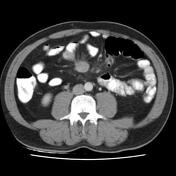
Stercoral collitis

Published
29 Mar 2023
77% complete
X-ray
CT
Case
Post-primary pulmonary tuberculosis

Published
15 Mar 2013
68% complete
CT
Case
Loculated tension hydropneumothorax

Published
24 May 2023
80% complete
X-ray
CT
Case
Klebsiella pneumoniae necrotising fasciitis - post intramuscular injection

Published
28 Jun 2023
80% complete
X-ray
CT
Annotated image
Case
Mesenteric neuroendocrine tumour metastasis
Published
07 Jun 2022
95% complete
CT
Article
Sphincter pupillae muscle
The sphincter pupillae muscle is a circular ring of smooth muscle within the iris responsible for constriction of the pupil (miosis). The structure is stimulated by the parasympathetic nervous system causing the muscle to decrease in diameter as it contracts.
Gross anatomy
The sphincter pupill...
Case
Pineal cyst

Published
07 Jan 2021
74% complete
CT
Case
Meningitis from mastoiditis resulting in death

Published
05 Sep 2010
89% complete
CT
MRI
DSA (angiography)
Article
Juxtaphrenic peak sign
The juxtaphrenic peak sign, also known as diaphragmatic tenting or Kattan sign, refers to the peaked or tented appearance of a hemidiaphragm which can occur in the setting of lobar collapse or post lobectomy (lung). It is caused by retraction of the lower end of diaphragm at an inferior accessor...
Case
Petroclival meningioma

Published
02 Nov 2014
77% complete
Annotated image
MRI
Case
Foreign body in stomach

Published
29 Dec 2022
91% complete
X-ray
Article
Colonic atresia
Colonic atresia is a rare congenital malformation in which parts of colon are absent. Contrary to anal atresia, the anal opening is present. Multiple atretic colonic segments may occur simultaneously.
Radiographic features
Fluoroscopy
Contrast enema typically shows no passage of contrast prox...
Case
Diabetic foot

Published
08 Apr 2022
75% complete
X-ray


 Unable to process the form. Check for errors and try again.