


308 results
Article
Urethral duplication
Urethral duplication is a rare condition in which either a part of the entire urethra is duplicated. It usually occurs in the sagittal plane, and the more dorsal copy is usually the duplication.
Pathology
Urethral duplications may occur due to a variety of developmental anomalies. In females, ...
Article
Lymphangioleiomyomatosis
Lymphangioleiomyomatosis (LAM) is a low-grade destructive metastasising PEComatous tumour 1 resulting from the proliferation of LAM cells in the lung, kidney and axial lymphatics. The disease is caused by mutations of the TSC2 or TSC1 genes and is more commonly sporadic rather than inherited. Cy...
Case
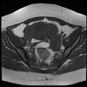
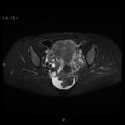
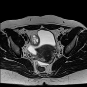
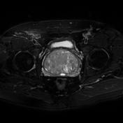
Uterine leiomyomas and bilateral ovarian fibromas
Published
13 Apr 2019
68% complete
MRI
Case
Avascular necrosis - bilateral femoral heads
Published
19 Nov 2020
95% complete
MRI
CT
Article
Deep vein thrombosis
Deep vein thrombosis (DVT) most commonly occurs in the lower limbs, however, are not uncommon in the upper limb and neck deep veins. Other types of venous thrombosis, such as intra-abdominal and intracranial, are discussed in separate articles.
Terminology
The term indeterminate (equivocal) DV...
Article
Rectus femoris muscle injury
Rectus femoris muscle injuries are muscle injuries, which encompass contusions, strains, tears and avulsions of the rectus femoris muscle.
Epidemiology
Rectus femoris muscle injuries are a common injury in athletes, especially in football/soccer players 1. The rectus femoris muscle is most fr...
Article
Ovarian serous cystadenoma
Ovarian serous cystadenomas are a type of benign ovarian epithelial tumour at the benign end of the spectrum of ovarian serous tumours.
Terminology
Serous ovarian tumours are traditionally described with a "cyst-" prefix because of their primarily cystic composition, e.g. cystadenoma, cystaden...
Article
Coccydynia
Coccydynia refers to pain in and among the area of the coccyx. It is characterised by coccygeal pain which is typically provocated by pressure. It may remain unclear in origin owing to the unpredictability of the source of pain 1.
Epidemiology
No accurate data about the frequency of coccydynia...
Article
Labelled imaging anatomy cases
This article lists a series of labelled imaging anatomy cases by body region and modality.
Brain
CT head: non-contrast axial
CT head: non-contrast coronal
CT head: non-contrast sagittal
CT head: non-contrast axial with clinical questions
CT head: angiogram axial
CT head: angiogram coronal...
Case
Uterine adenomyosis
Published
05 May 2015
59% complete
MRI
Article
Pelvic insufficiency fractures
Pelvic insufficiency fractures are a relatively common subtype of insufficiency fracture, and are recognised as a major cause of low back, buttock and groin pain in susceptible populations.
Epidemiology
Senile/postmenopausal osteoporosis is the most common predisposing factor. Other important ...
Case
Perianal fistula - inter-sphincteric type
Published
02 Nov 2018
68% complete
MRI
Article
Colorectal cancer (summary)
This is a basic article for medical students and other non-radiologists
Colorectal cancer, also called colorectal carcinoma (CRC), is the most common cancer of the gastrointestinal tract and the second most frequently diagnosed malignancy in adults. CT and MRI are the modalities most frequently...
Article
Haemophilic pseudotumour
Haemophilic pseudotumours are rare complications of haemophilia consisting of a progressive cystic swelling of muscle and/or bone due to repeated bleeding, occurring in <2% of haemophiliacs.
Epidemiology
Haemophilic pseudotumours are reported in 1-2% of patients with haemophilia.
Clinical pr...
Case
Non Hodgkin's lymphoma

Published
08 Mar 2021
95% complete
CT
MRI
Case
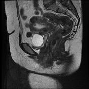
Isolated fallopian tube torsion with a paratubal cyst
Published
03 Apr 2018
92% complete
MRI
Article
Pelvic phlebography
Pelvic phlebography or pelvic venography is an interventional procedure, in which the pelvic and gonadal veins are opacified to assess venous and collateral system anatomy for the diagnosis, treatment and pre-operative planning of pelvic vein pathology.
This procedure is usually performed at th...
Article
Iliofemoral deep vein thrombosis
Iliofemoral deep vein thrombosis (DVT) occurs when a thrombus in the iliac vein (common, external or internal) and/or common femoral vein obstructs the venous outflow from the lower limb leading to marked oedema. DVT of the IVC or the more distal lower limb veins may also be present.
Terminolog...
Case
Benign prostatic nodular hyperplasia
Published
29 Feb 2012
45% complete
MRI
Playlist
August 2013


10 cases
Use the following history (from recalls on RANZCR website). P.S. cases 2 and 3 are part of one case. Question 1 73F, long hx of COPD...


 Unable to process the form. Check for errors and try again.