
Search results for “( "Lateral Elbow Tendinopathy" OR Tennis+Elbow OR Lateral+Epicondylitis)”
Did you mean
dorso-lateral elbow tendinopathy or?


85 results found
Article
Lateral humeral condyle fracture
Lateral humeral condyle fractures also referred to simply as lateral condyle fractures (in the appropriate context), are relatively common elbow fractures that predominantly occur in children. They may be subtle but are hugely important to diagnose promptly because if they are missed, they tend ...
Case
Lateral epicondyle avulsion fracture

Published
08 Aug 2010
100% complete
Annotated image
X-ray
Article
Radius
The radius (plural: radii) is one of the two long bones present in the forearm, located laterally in the supinated anatomical position. It has a smaller proximal end and enlarges to a larger distal end (opposite to the ulna).
Gross anatomy
Osteology
The proximal radius comprises the articula...
Article
Humerus
The humerus (plural: humeri) is a tubular bone of the arm that articulates proximally at the shoulder with the glenoid of the scapula, and distally at the elbow, with the radius and ulna.
Gross anatomy
Osteology
The humerus begins proximally as a rounded head and joins the greater and lesser ...
Article
Elbow (lateral view)
The lateral elbow view is part of the two view elbow series, examining the distal humerus, proximal radius and ulna. It is deceptively one of the more technically demanding projections in radiography 1-3.
The projection is the orthogonal view of the AP elbow allowing for examination of the ulna...
Case
Lateral epicondyle fracture (elbow)
Published
03 Jun 2020
92% complete
X-ray
CT
Case
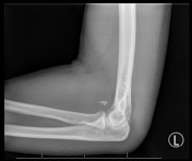
Lateral epicondylitis
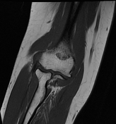
Published
03 Feb 2024
92% complete
MRI
Article
Lateral epicondyle fracture (elbow)
Lateral epicondyle fractures of the elbow are rare epicondylar fractures. They are much rarer than medial epicondyle fractures and represent avulsion of the lateral epicondyle. They are usually seen in the setting of other injuries 1-3.
Terminology
These fractures are avulsion fractions of th...
Article
Posterolateral rotatory instability of the elbow
Posterolateral rotatory instability (PLRI) of the elbow is the most common pattern of elbow instability, most commonly seen following posterior elbow dislocation 1. It is characterized by 2-4:
posterolateral subluxation/dislocation of the radial head relative to the capitellum
posterior displa...
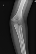
Case
Lateral epicondylitis

Published
08 Jan 2024
68% complete
X-ray
Ultrasound
Article
Ossification centers of the elbow
There are six ossification centers of the elbow that appear and develop in a relatively reproducible fashion, and are key to assessment of the pediatric elbow radiograph. Timing of their appearance varies in the literature but an approximation is given below. A useful mnemonic to remember the or...
Article
Elbow ossification (mnemonic)
Mnemonics for elbow ossification include CRITOE and CRITOL. These are essentially the same, apart from the terminal letter which represents the External or Lateral epicondyle.
Mnemonics
CRITOE
C: capitellum
R: radial head
I: internal epicondyle
T: trochlea
O: olecranon
E: external epicon...
Article
Elbow ossification
Elbow ossification occurs at the six elbow ossification centers in a reproducible order. Being familiar with the order of ossification of the elbow is important in not mistaking an epicondylar fracture for a normal ossification center.
Appearance
Order
The order of appearances of the elbow o...
Case
Salter-Harris type II fracture of proximal radius
Published
22 Feb 2021
97% complete
X-ray
Annotated image
Case
Lateral epicondylitis

Published
18 Jun 2014
79% complete
Ultrasound
Case
Acute calcific periarthritis (elbow)

Published
03 Jul 2020
79% complete
X-ray
Ultrasound
Case
Extensor tendinosis - elbow

Published
28 Apr 2016
63% complete
Ultrasound
Article
Anconeus muscle
The anconeus muscle is a small muscle in the posterior compartment of the arm at the lateral aspect of the elbow. Its functional significance is not well understood 5.
It should not be confused with the anconeus epitrochlearis, an accessory muscle that is present in up to one third of people, a...
Article
Knee joint
The knee joint is a modified hinge joint between the femur, tibia, and patella. It is the largest synovial joint in the body and allows flexion and extension of the leg as well as some rotation in the flexed position.
Summary
location: two condylar joints between femur and tibia; saddle joint ...
Case
Fracture of capitellum and humeral trochlea - Mckee double arc sign

Published
19 Feb 2023
75% complete
X-ray
Annotated image


 Unable to process the form. Check for errors and try again.