

Search results for “chest”


204 results
Article
CT chest abdomen-pelvis (protocol)
The CT chest-abdomen-pelvis protocol serves as an outline for an examination of the trunk covering the chest, abdomen and pelvis. It is one of the most common CT examinations conducted in routine and emergencies. It can be combined with a CT angiogram.
Note: This article aims to frame a genera...
Case
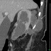
Ruptured renal angiomyolipoma causing pleuritic chest pain diagnosed on CTPA

Published
08 Oct 2023
92% complete
DSA (angiography)
CT
Case
Perinephric abscess pointing into the posterior chest wall
Published
28 Nov 2015
80% complete
CT
Case
Renal stone (chest X-ray)

Published
26 May 2023
77% complete
X-ray
Case
Renal cell carcinoma - incidental on chest CT

Published
26 Jun 2019
65% complete
CT
Case
Normal dual phase CT chest, abdomen and pelvis (trauma protocol)

Published
23 Mar 2023
65% complete
CT
Question
Question 1687
A 60-year-old presents with cough and has a chest radiograph. Which review area is the region of abnormality? [Frontal chest radiograph]
Question
Question 2212
A 20-year-old woman presents with a 6-month history of dry cough and chest pain. Based on the lateral chest X-ray, what is the most likely diagnosis?
Question
Question 2213
A 30-year-old woman presents with a 6-month history of dry cough and chest pain. Based on the lateral chest X-ray, what is the most likely diagnosis?
Question
Question 814
Which radiologic sign describes the pathology on this chest radiograph?
Article
Whole-body CT (protocol)
CT polytrauma/multitrauma, also called trauma CT, whole body CT (WBCT) or panscan, is an increasingly used investigation in patients with multiple injuries sustained after significant trauma.
The majority of the evidence regarding whole-body CT is, understandably, retrospective. There is some e...
Article
Labeled imaging anatomy cases
This article lists a series of labeled imaging anatomy cases by body region and modality.
Brain
CT head: non-contrast axial
CT head: non-contrast coronal
CT head: non-contrast sagittal
CT head: non-contrast axial with clinical questions
CT head: angiogram axial
CT head: angiogram coronal
...
Article
Urinothorax
Urinothorax (plural: urinothoraces), also known as urothorax, is a rare cause of pleural effusion due to the accumulation of urine within the pleural space.
Clinical presentation
Patients present with varying degrees of respiratory distress depending on the amount of fluid that has accumulated...
Article
Brachytherapy seed migration to the lung
Brachytherapy seed migration to the lung is a known complication of radioactive seed therapy. These seeds are used for localized treatment of malignancies, most commonly prostate cancer.
Regarding staging, nearly 79% of the cases are localized, 12% are regional and 5% present with distant disea...
Article
Milk of calcium (disambiguation)
The term milk of calcium (MOC) is given to dependent, sedimented calcification within a cystic structure or hollow organ. This sort of colloidal calcium suspension layering can occur in various regions:
renal: milk of calcium in renal cyst (most common)
ureter: milk of calcium in the ureter 7
...
Article
Body imaging
Body imaging is the term assigned to cross-sectional imaging of the body, which radiologically refers to the chest, abdomen and pelvis. It is often used by radiologists who report this region (sometimes known as body imagers/radiologists) to differentiate their primary area of interest from othe...
Question
Question 691
A 70-year-old has long-standing cutaneous lesions, as shown on the accompanying axial CT of the chest. What is the most likely underlying condition?
Article
Viscera
The viscera (singular: viscus) refers to all the internal organs within the major cavities of the thorax, abdomen and pelvis. Therefore it does not include organs of the CNS, head and neck or musculoskeletal compartments nor does it encompass non-internal organs (e.g. the skin) 1.
Splanchnology...
Article
Sickle cell disease
Sickle cell disease (SCD) (historically also known as drepanocytosis) is a hereditary (autosomal recessive) condition resulting in the formation of abnormal hemoglobin (a hemoglobinopathy), which manifests as multisystem ischemia and infarction, as well as hemolytic anemia.
Hemoglobin SC (HbSC...
Article
Normal imaging examples
This article lists examples of normal imaging divided by body region and system.
brain
head and neck
spine
chest
breast
gastrointestinal
genitourinary
hepatobiliary
upper limb
lower limb
pediatrics


 Unable to process the form. Check for errors and try again.