


21,832 results
Case
Intraosseous lipoma

Published
13 Apr 2011
82% complete
X-ray
Case
Os subepicondylare mediale

Published
27 May 2022
72% complete
X-ray
Case
Gynecomastia

Published
13 Apr 2011
66% complete
Ultrasound
Case
Non-accidental injury - skull fractures

Published
15 Nov 2022
80% complete
CT
Case
Os supranaviculare
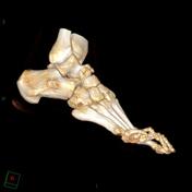
Published
31 Oct 2011
71% complete
CT
Case
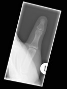
Finger PIP joint septic arthritis and osteomyelitis

Published
01 Sep 2020
94% complete
X-ray
Article
Mid-talar axis
The mid-talar axis represents a line drawn down the longitudinal axis of the talus and can be drawn on lateral and DP radiographs.
Measurement
Independent on the view on which the line is drawn, it should bisect the neck of the talus and the head.
On the lateral and DP views, the line should...
Case
Achilles tendon injury

Published
02 May 2014
66% complete
X-ray
Ultrasound
Case
PASTA Lesion

Published
14 Mar 2018
89% complete
MRI
Case
Stieda fracture

Published
19 Jan 2022
91% complete
X-ray
Case
Acromioclavicular joint ganglion and long head of biceps brachii dislocation

Published
06 Feb 2011
63% complete
Ultrasound
Case
Gamekeeper thumb
Published
24 Jan 2018
91% complete
X-ray
Case
Parameniscal cyst

Published
09 Jul 2022
68% complete
X-ray
MRI
Case
Metacarpal fracture

Published
18 Jan 2022
94% complete
X-ray
Case
Right pubic rami fractures

Published
16 Oct 2014
91% complete
X-ray
Article
Lateral talocalcaneal angle
The lateral talocalcaneal angle is one of the angles that can be measured for the assessment of pes planus and pes cavus and assessment of hindfoot deformity.
Measurement
The lateral talocalcaneal angle is drawn on a weight-bearing lateral foot radiograph. There are two ways that it has been d...
Case
Calcaneofibular ligament bony avulsion

Published
07 May 2023
91% complete
X-ray
Ultrasound
Case
Anterior shoulder dislocation

Published
09 Jan 2017
88% complete
X-ray
Case
Rolando fracture

Published
19 Nov 2023
94% complete
X-ray
Article
Chordoma
Chordomas are uncommon malignant tumors of the axial skeleton that account for 1% of intracranial tumors and 4% of all primary bone tumors.
They originate from embryonic remnants of the primitive notochord (earliest fetal axial skeleton, extending from the Rathke's pouch to the tip of the cocc...


 Unable to process the form. Check for errors and try again.