


121,153 results
Case
Intraosseous lipoma

Published
13 Apr 2011
82% complete
X-ray
Case
Cystic hygroma

Published
22 Jan 2022
92% complete
CT
X-ray
Ultrasound
Photo
Case
Breast ductal carcinoma in situ

Published
12 Feb 2018
75% complete
Mammography
Ultrasound
Case
Os subepicondylare mediale

Published
27 May 2022
72% complete
X-ray
Case
Gynecomastia

Published
13 Apr 2011
66% complete
Ultrasound
Case
Peritoneal inclusion cyst

Published
05 Feb 2011
66% complete
Ultrasound
Case
Bilateral hydrosalpinx

Published
09 Aug 2021
82% complete
Fluoroscopy
Article
Kaposiform hemangioendothelioma
Kaposiform hemangioendotheliomas are rare, locally invasive vascular tumors that often present in infancy, most commonly as an enlarging cutaneous mass 1,2. They are classified as distinct from tufted angiomas in the ISSVA classification of vascular anomalies. However, some consider it to be on ...
Case
Non-accidental injury - skull fractures

Published
15 Nov 2022
80% complete
CT
Case
Marginal cord insertion

Published
29 Sep 2023
97% complete
Ultrasound
Case
Os supranaviculare
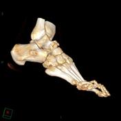
Published
31 Oct 2011
71% complete
CT
Case
Mesothelioma - subtle nodular thickening of the interlobar fissures

Published
09 May 2018
92% complete
CT
Annotated image
X-ray
Case
Finger PIP joint septic arthritis and osteomyelitis

Published
01 Sep 2020
94% complete
X-ray
Article
Mid-talar axis
The mid-talar axis represents a line drawn down the longitudinal axis of the talus and can be drawn on lateral and DP radiographs.
Measurement
Independent on the view on which the line is drawn, it should bisect the neck of the talus and the head.
On the lateral and DP views, the line should...
Case
Achilles tendon injury

Published
02 May 2014
66% complete
X-ray
Ultrasound
Case
Tongue stud

Published
01 Jan 2009
59% complete
CT
Case
PASTA Lesion

Published
14 Mar 2018
89% complete
MRI
Case
Gastric carcinoma

Published
27 Jul 2014
71% complete
CT
Case
Stieda fracture

Published
19 Jan 2022
91% complete
X-ray
Case
Steinstrausse - multilevel

Published
20 May 2016
92% complete
CT


 Unable to process the form. Check for errors and try again.