
Items tagged “msk”
54 results
Case
Cysticercosis - soft tissue

Published
17 Apr 2015
91% complete
X-ray
Case
Inferior shoulder dislocation

Published
10 Oct 2015
97% complete
X-ray
Case
Scurvy

Published
27 Oct 2015
89% complete
X-ray
MRI
Case
Longitudinal epiphyseal bracket

Published
05 Nov 2015
86% complete
X-ray
MRI
Case
Fibroadipose vascular anomaly

Published
27 Oct 2015
89% complete
MRI
Case
Giant cell tumor of bone - maxilla

Published
29 Oct 2015
95% complete
CT
Case
Gollop-Wolfgang complex

Published
08 Nov 2015
75% complete
X-ray
Pathology
Article
Hands
The hand is part of the upper limb below the forearm and wrist. In the supinated anatomical position, the palm is facing anteriorly and the dorsum posteriorly.
The bones of the hand are:
carpals (8)
scaphoid
lunate
triquetrum
pisiform
trapezium
trapezoid
capitate
hamate
metacarpals (5...
Case
Calcific bursitis

Published
12 Jan 2016
98% complete
X-ray
MRI
Case
April Fools' 2016: Cactus disease (paleo-induced mineral periostitis)

Published
29 Mar 2016
88% complete
Photo
X-ray
Case
Monteggia fracture-dislocation

Published
08 Apr 2016
41% complete
X-ray
Case
Paget disease of femur

Published
12 May 2016
75% complete
MRI
CT
Case
Osteochondroma - vertebral

Published
01 Jun 2016
75% complete
MRI
CT
Case
First metatarsophalangeal joint anatomy

Published
16 May 2016
22% complete
Diagram
Case
Fracture through congenitally fused 5th phalanges

Published
30 Jul 2016
85% complete
X-ray
Annotated image
Case
Facet joint dislocation, Jefferson and hangman fractures

Published
26 Oct 2016
95% complete
X-ray
CT
MRI
Article
Peritalar dislocation
Peritalar dislocation, also referred to as subtalar dislocation, involves the combined dislocation of the talocalcaneal/subtalar and talonavicular joints without the involvement of the tibiotalar and calcaneonavicular joints. It is generally associated with high-energy trauma and accounts for a ...
Case
Fracture healing diagrams
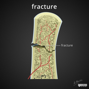
Published
23 Dec 2017
30% complete
Diagram
Case
Osteochondroma - scapular

Published
28 Jun 2018
93% complete
X-ray
Annotated image
Case
Transient synovitis - ankle

Published
20 Oct 2018
97% complete
X-ray


 Unable to process the form. Check for errors and try again.