
Items tagged “variant”
428 results
Case
Variant renal arteries

Published
04 Jun 2017
56% complete
CT
Case
Common celiacomesentric trunk

Published
27 Jun 2017
95% complete
CT
Case
Retropharyngeal carotid arteries (kissing carotids)

Published
02 Jul 2017
95% complete
CT
Annotated image
Case
Bilateral accessory parotid glands

Published
30 Jun 2017
92% complete
CT
Annotated image
Case
Kimerle anomaly

Published
13 Jul 2017
79% complete
X-ray
Case
Internal carotid artery aplasia/hypoplasia

Published
09 Sep 2017
71% complete
MRI
Article
Third condyle
The third condyle, also known as condylus tertius or the median occipital condyle, is a rare anatomical variant of the occipital bone that may mimic an occipital condyle fracture. It is part of the spectrum of occipital vertebrae.
Epidemiology
The third condyle is a rare variant, found in appr...
Case
Partial anomalous pulmonary venous return

Published
03 Dec 2017
89% complete
CT
Case
Azygos continuation of the inferior vena cava

Published
06 Dec 2017
84% complete
CT
Case
Os intermetatarseum

Published
12 Dec 2017
88% complete
X-ray
Case
Os patella cubiti

Published
19 Mar 2018
57% complete
Ultrasound
X-ray
Case
Accessory sacroiliac joints
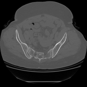
Published
13 Feb 2018
92% complete
CT
Case
Fenestration of vertebral artery

Published
13 Feb 2018
74% complete
CT
Article
Accessory sacroiliac joint
Accessory sacroiliac joints are a common finding, present on ~15% (range 13-17.5%) of CT studies, and may be unilateral or bilateral. They are an articulation between the medial aspect of the posterior superior iliac spine and the sacrum just lateral to the second dorsal sacral foramen. They may...
Case
Hypoplastic right 12th rib

Published
26 Feb 2018
74% complete
Annotated image
CT
X-ray
Article
Accessory semimembranosus muscle
The accessory semimembranosus muscle is a rare accessory muscle of the posterior compartment of the thigh. It arises from the distal aspect of the semimembranosus muscle belly and courses through the popliteal fossa between it and the semitendinosus muscle medially and the biceps femoris lateral...
Case
Tilted sternum

Published
02 Apr 2018
92% complete
CT
Case
Subseptate uterus

Published
05 Apr 2018
92% complete
MRI
Case
Retroaortic left renal vein - type II

Published
17 Apr 2018
92% complete
CT
Case
Duplicated inferior vena cava

Published
30 Apr 2018
98% complete
CT


 Unable to process the form. Check for errors and try again.