
The anteroposterior elbow view for pediatrics is part of the two view elbow series, examining the distal humerus, proximal radius and ulna.
On this page:
Indications
The projection demonstrates the elbow joint in its natural anatomical position allowing for adequate radiographic examination of the articulations of the elbow including the radiohumeral and humeroulnar joints.
It is useful in pediatric imaging for evaluating infections, suspected dislocations or fractures, and localizing foreign bodies in the distal humerus, and proximal forearm.
Patient position
- patient is seated next to the table
- the fully extended arm and forearm, in a supinated position, are kept in contact with the tabletop
- ensure all aspects of the arm from the wrist to the humerus are in the same plane
Technical factors
- anteroposterior projection
-
centering point
- midpoint between the humeral epicondyles
-
collimation
- superior to the distal third of the humerus
- inferior to include one-third of the proximal radius and ulna
- lateral to include the skin margin
- medial to include medial skin margin
-
orientation
- portrait
-
detector size
- 18 cm x 24 cm
-
exposure 1
- 50-57 kVp
- 2-3 mAs
-
SID
- 110 cm
-
grid
- no
Image technical evaluation
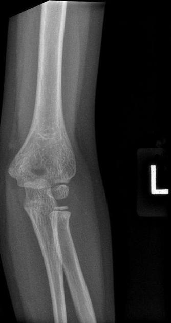
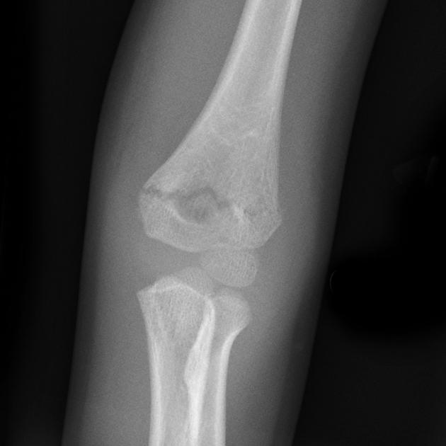
- the elbow is in an AP position, with slight internal rotation.
- patient's arm should be rotated externally to ensure that the trochlea and capitellum are seen in profile.
Practical points
Preparing the room beforehand (setting up the detector, exposure and preparing lead gowns) is extremely beneficial for elbow imaging as young children may begin to cry the moment their affected arm is brought away from their body.
For patients who are unable to properly extend their elbow, partial flexion may be used; at initial presentation, two AP views may then be required:
- one with the table top lowered and the forearm flat against the detector
- another with the table top raised and the humerus flat against the detector, using an immobilization sponge to elevate the distal forearm
Immobilization techniques
To prevent malrotation/motion artifact in the radiograph, parental holding at the proximal half of the child’s arm and distal half of the forearm may be required.
- if the parent is accompanying the child, whilst the parent puts on a lead gown, it is the radiographer's responsibility to ensure the child does not fall off the chair
- other alternative methods such as distraction techniques may be ideal to avoid scattered radiation to parents and staff 2


 Unable to process the form. Check for errors and try again.