
Double outlet right ventricle (DORV) is a congenital cardiac anomaly where both the aorta and pulmonary trunk arise from the morphologically right ventricle. It is reported to account for ~2% of congenital cardiac defects 1. It is usually classed as a conotruncal anomaly. There is almost always a concurrent ventricular septal defect (VSD) 4.
On this page:
Epidemiology
The estimated incidence is at ~1:10,000 births.
Associations
-
aneuploidic/chromosomal
- trisomy 13 1
- trisomy 18 1
-
cardiovascular/pulmonary
- congenital pulmonary stenosis
- coarctation of the aorta
- right-sided aortic arch
- anomalous pulmonary venous return
- other
Pathology
Genetics
Most cases are thought to have a sporadic occurrence.
Types
According to the position of the great vessels 5:
- side by side positioning of great vessels
- right-sided malpositioning of great vessels
- left-sided malpositioning of great vessels
According to where the VSD is located about the great vessels 6:
- DORV with subaortic VSD
- VSD is located just below the aorta
- DORV with subpulmonary VSD (also called Taussig-Bing anomaly)
- VSD is located below the pulmonary artery
- DORV with doubly committed VSD
- VSD under both of the great arteries
- DORV with non-committed (or remote) VSD
- VSD is not located near the aorta or the pulmonary artery
Radiographic features
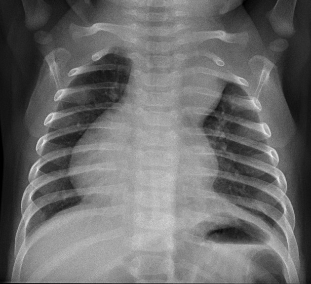
Plain radiograph
Appearances on chest radiographs are variable depending on the subclassification and presence of concurrent other anomalies. May show evidence of right ventricular enlargement.
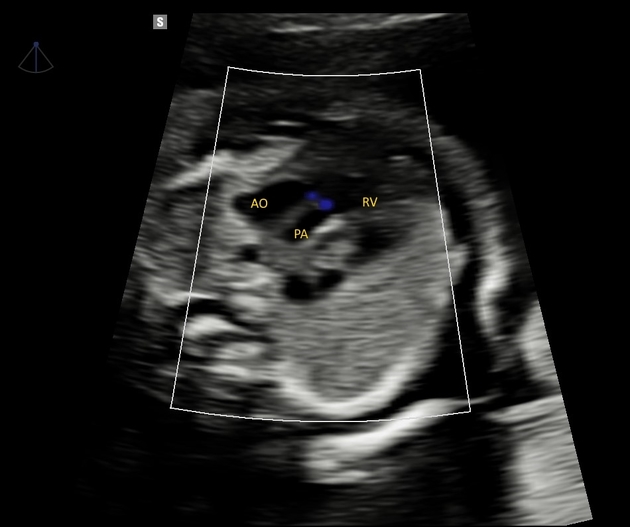
Ultrasound
On echocardiography, there is typically a lack of communication between the posterior aortic root and the anterior mitral valve leaflet.
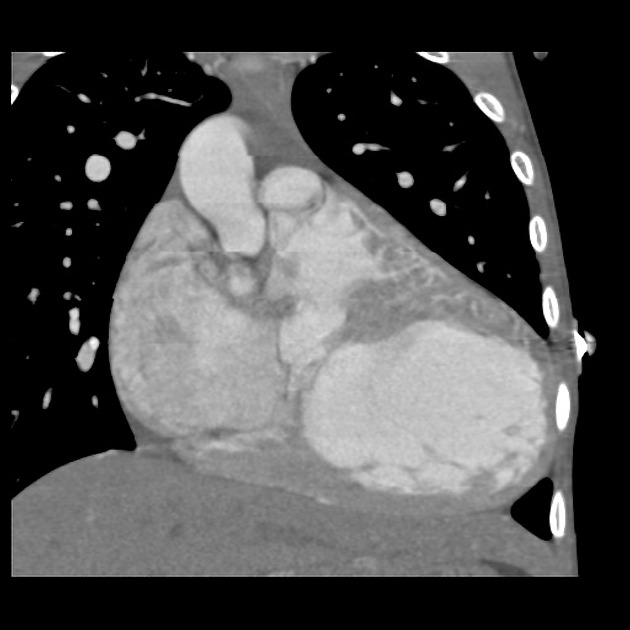
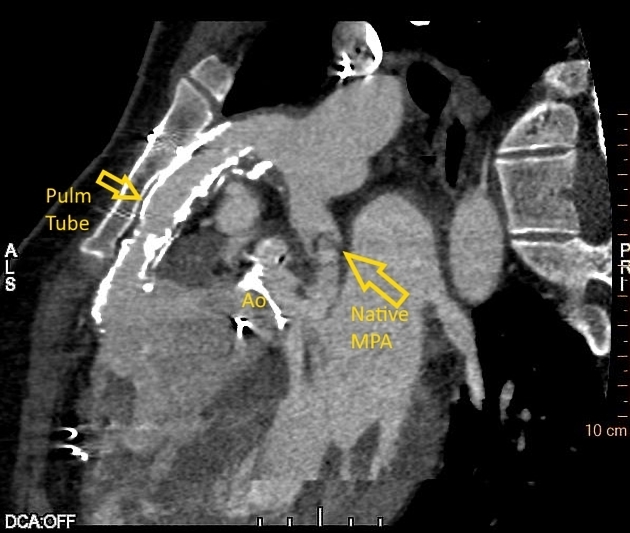
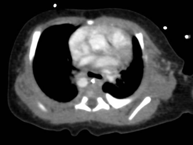
CT/MRI
Allows direct visualization of abnormal anatomy.


 Unable to process the form. Check for errors and try again.