
Frontal sinus
Citation, DOI, disclosures and article data
At the time the article was created Frank Gaillard had no recorded disclosures.
View Frank Gaillard's current disclosuresAt the time the article was last revised Tariq Walizai had no financial relationships to ineligible companies to disclose.
View Tariq Walizai's current disclosures- Frontal sinuses
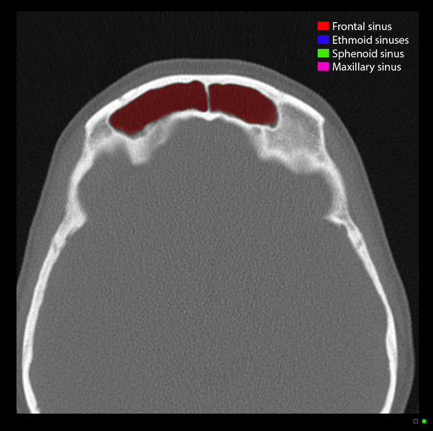
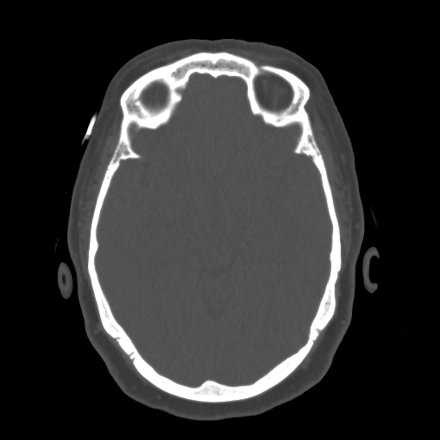
The frontal sinuses are the paranasal sinuses within the frontal bone. They are lined with mucosa and are most often two in number.
On this page:
Summary
location: anterior frontal bones on either side of the midline behind the brow ridges
blood supply: supratrochlear, supraorbital and anterior ethmoidal arteries
innervation: supraorbital and supratrochlear nerves
Gross anatomy
The frontal sinus has two chambers, one on each side, and they are almost always asymmetrical and separated by a septum. Each sinus extends superior to the medial end of the eyebrow and back into the orbital portion of the frontal bone. However, three or more chambers may be present in ~10% (range 1.5%-21%). It is divided by thin bony intrasinus septa, usually off-midline and rarely dehiscent.
The orbit and anterior cranial fossa form important relations to these sinuses.
Drainage from the frontal sinus tends to be more variable than the other paranasal sinuses and there is inconsistent terminology used in its anatomic description 3. In general, the frontal sinus outflow tract consists of a narrowing at the lower medial corner of the sinus (frontal infundibulum), where an ostium (opening) is demarcated by a small ridge of bone at the anterior sinus wall. Inferior to the ostium, drainage continues along a narrow passage known as the frontal recess 4 or superior compartment of the frontal sinus drainage pathway 3. At this point, variant anatomy of the frontal/ethmoidal bone junction leads to two main variations:
drainage into ethmoidal infundibulum, through the hiatus semilunaris into the middle meatus
drainage directly into middle meatus
ADVERTISEMENT: Supporters see fewer/no ads
Arterial supply
The frontal sinus is supplied by the supratrochlear, anterior ethmoidal, and supraorbital arteries, all of which are branches of the ophthalmic artery.
Venous drainage
Venous drainage is via the superior ophthalmic veins.
Lymphatic drainage
Lymph drainage of the frontal sinus is into the submandibular nodes (cf. the overlying skin which drains to the preauricular group of nodes).
Variant anatomy
-
may be absent (i.e. aplasia) or underdeveloped (i.e. hypoplasia): unilateral (4%) or bilateral (5%)
this may be associated with a metopic suture
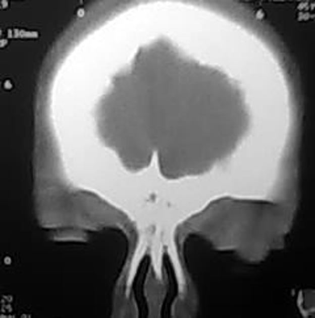
may be large: extending through zygomatic processes, orbital bones, and into the squamae
Development
Frontal sinus begin as an outpouching of the lateral nasal wall, at the level of middle nasal meatus. This outpouching then extends superomedially giving origin to the ethmoidal cells and the frontal recess. The frontal recess then pneumatized into the frontal bone 5. Development begins late in intrauterine life (at 3 to 4 months) 5. However, frontal sinus are not present a birth 6. Pneumatization develops from 1-2 years old 5.
References
- 1. Danesh-Sani S, Bavandi R, Esmaili M. Frontal Sinus Agenesis Using Computed Tomography. J Craniofac Surg. 2011;22(6):e48-51. doi:10.1097/SCS.0b013e318231e26c - Pubmed
- 2. Mcminn. Last's Anatomy. (2003) ISBN: 9780729537520 - Google Books
- 3. Daniels D, Mafee M, Smith M et al. The Frontal Sinus Drainage Pathway and Related Structures. AJNR Am J Neuroradiol. 2003;24(8):1618-27. PMC7973969 - Pubmed
- 4. Huang B, Lloyd K, DelGaudio J, Jablonowski E, Hudgins P. Failed Endoscopic Sinus Surgery: Spectrum of CT Findings in the Frontal Recess. Radiographics. 2009;29(1):177-95. doi:10.1148/rg.291085118 - Pubmed
- 5. Lee S, Fernandez J, Mirjalili S, Kirkpatrick J. Pediatric Paranasal Sinuses—Development, Growth, Pathology, & Functional Endoscopic Sinus Surgery. Clin Anat. 2022;35(6):745-61. doi:10.1002/ca.23888 - Pubmed
- 6. Jacob S. Head and Neck. Human Anatomy. 2008;:181-225. doi:10.1016/b978-0-443-10373-5.50010-5
Incoming Links
- Frontalis muscle
- Metopic suture
- Middle meatus
- Patterns of sinonasal obstruction
- Depressed skull fracture
- Hiatus semilunaris
- Anterior ethmoidal artery
- Paranasal sinus mucocele
- Paranasal sinuses and facial bones (lateral view)
- Frontal sinus fracture
- Allergic fungal sinusitis
- Frontal bullar cells
- Skull (Caldwell view)
- Supra agger cell
- Kuhn classification
- Frontal infundibulum
- Frontal bone
- Supraorbital air cells
- Maxillary sinus carcinoma (staging)
- Ethmoid infundibulum
- Orbit roof subperiosteal collection
- Persistent metopic suture with frontal sinus agensis
- Frontal sinus osteoma
- Pott puffy tumor
- Paranasal sinus development (Gray's illustration)
- Esthesioneuroblastoma
- Frontal sinus osteoma
- Dental abscess
- Persistent metopic suture
- Frontal sinuses aplasia
- Persistent metopic suture
- Sinonasal angiomatous polyp
- Orbital metastasis of breast cancer
- Frontoethmoidal sinus osteoma
- Giant frontal sinus osteoma
- Frontoethmoidal mucocele
- Frontal sinus mucocele
- Concomitant orbital blow-in and blow-out fractures
- Frontal sinus osteoma
- Frontal mucocele and sinonasal polyposis
Related articles: Anatomy: Head and neck
- skeleton of the head and neck
-
cranial vault
- scalp (mnemonic)
- fontanelle
-
sutures
- calvarial
- facial
- frontozygomatic suture
- frontomaxillary suture
- frontolacrimal suture
- frontonasal suture
- temporozygomatic suture
- zygomaticomaxillary suture
- parietotemporal suture (parietomastoid suture)
- occipitotemporal suture (occipitomastoid suture)
- sphenofrontal suture
- sphenozygomatic suture
- spheno-occipital suture (not a true suture)
- lacrimomaxillary suture
- nasomaxillary suture
- internasal suture
- basal/internal
- skull landmarks
- frontal bone
- temporal bone
- parietal bone
- occipital bone
- skull base (foramina)
-
facial bones
- midline single bones
- paired bilateral bones
- cervical spine
- hyoid bone
- laryngeal cartilages
-
cranial vault
- muscles of the head and neck
- muscles of the tongue (mnemonic)
- muscles of mastication
-
facial muscles
- epicranius muscle
- circumorbital and palpebral muscles
- nasal muscles
-
buccolabial muscles
- elevators, retractors and evertors of the upper lip
- levator labii superioris alaeque nasalis muscle
- levator labii superioris muscle
- zygomaticus major muscle
- zygomaticus minor muscle
- levator anguli oris muscle
- malaris muscle
- risorius muscle
- depressors, retractors and evertors of the lower lip
- depressor labii inferioris muscle
- depressor anguli oris muscle
- mentalis muscle
- compound sphincter
-
orbicularis oris muscle
- incisivus labii superioris muscle
- incisivus labii inferioris muscle
-
orbicularis oris muscle
- muscle of mastication
- modiolus
- elevators, retractors and evertors of the upper lip
- muscles of the middle ear
- orbital muscles
- muscles of the soft palate
- pharyngeal muscles
- suprahyoid muscles
- infrahyoid muscles
- intrinsic muscles of the larynx
- muscles of the neck
- platysma muscle
- longus colli muscle
- longus capitis muscle
- scalenus anterior muscle
- scalenus medius muscle
- scalenus posterior muscle
- scalenus pleuralis muscle
- sternocleidomastoid muscle
-
suboccipital muscles
- rectus capitis posterior major muscle
- rectus capitis posterior minor muscle
- obliquus capitis superior muscle
- obliquus capitis inferior muscle
- accessory muscles of the neck
- deep cervical fascia
-
deep spaces of the neck
- anterior cervical space
- buccal space
- carotid space
- danger space
- deep cervical fascia
- infratemporal fossa
- masticator space
- parapharyngeal space
- stylomandibular tunnel
- parotid space
- pharyngeal (superficial) mucosal space
- perivertebral space
- posterior cervical space
- pterygopalatine fossa
- retropharyngeal space
- suprasternal space (of Burns)
- visceral space
- surgical triangles of the neck
- orbit
- ear
- paranasal sinuses
- upper respiratory tract
- viscera of the neck
- blood supply of the head and neck
-
arterial supply
-
common carotid artery
- carotid body
- carotid bifurcation
- subclavian artery
- variants
-
common carotid artery
- venous drainage
-
arterial supply
- innervation of the head and neck
-
cranial nerves
- olfactory nerve (CN I)
- optic nerve (CN II)
- oculomotor nerve (CN III)
- trochlear nerve (CN IV)
-
trigeminal nerve (CN V) (mnemonic)
- trigeminal ganglion
- ophthalmic division
- maxillary division
- mandibular division
- abducens nerve (CN VI)
- facial nerve (CN VII)
-
vestibulocochlear nerve (CN VIII)
- vestibular ganglion (Scarpa's ganglion)
- glossopharyngeal nerve (CN IX)
- vagus nerve (CN X)
- (spinal) accessory nerve (CN XI)
- hypoglossal nerve (CN XII)
- parasympathetic ganglia of the head and neck
- cervical sympathetic ganglia
- greater occipital nerve
- third occipital nerve
-
cervical plexus
- muscular branches
- longus capitis
- longus colli
- scalenes
- geniohyoid
- thyrohyoid
-
ansa cervicalis
- omohyoid (superior and inferior bellies separately)
- sternothyroid
- sternohyoid
- phrenic nerve
- contribution to the accessory nerve (CN XI)
- cutaneous branches
- muscular branches
- brachial plexus
- pharyngeal plexus
-
cranial nerves
- lymphatic drainage of the head and neck
- embryological development of the head and neck


 Unable to process the form. Check for errors and try again.