
Olfactory nerve
Updates to Article Attributes
The olfactory nerve (cranial nerve I, a.k.a. CN I)and most rostral of the (CN I)cranial nervesnerve. Along with the optic nerve, it is actually a peripheral outpostextension of the central nervous system.
The bipolar cell is the first-order sensory neuronneurone located in the roof of the nasal cavity, immediately inferior to the cribriform plate of the ethmoid bone. This cell is analogous to the sensory cells of spinal nerves, whose cell bodies reside in the dorsal root ganglion. Their central processes form filaments (~20) which pass through the cribriform plate, pierce the dura mater and relay in the olfactory bulb, the expanded anterior portion of the nerve that sits in the olfactory fossa. The central process of these second-order neuronsneurones forms the olfactory tract which courses posteriorly, superior to the olfactory groove of the anterior cranial fossa, and inferior to the olfactory sulcus (lateral to gyrus rectus, and medial to the orbital gyri).
Anterior to the anterior perforated substance, they form medial and lateral olfactory striae. The lateral olfactory striae project to the uncus. The medial olfactory striae ultimately project to the hypothalamus and brainstem nuclei.
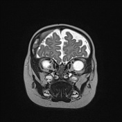
Coronal images are the best to depict the olfactory nerve as it is situated deep in the olfactory groove.
-<p>The <strong>olfactory nerve (cranial nerve I</strong>, a.k.a. <strong>CN I)</strong> is the first and most rostral of the <a href="/articles/cranial-nerves">cranial nerves</a>. Along with the <a href="/articles/optic-nerve">optic nerve</a>, it is actually a peripheral outpost of the <a title="Central nervous system" href="/articles/central-nervous-system-1">central nervous system</a>.</p><p>The bipolar cell is the first-order sensory neuron located in the roof of the <a href="/articles/nasal-cavity">nasal cavity</a>, immediately inferior to the <a href="/articles/cribriform-plate">cribriform plate</a> of the <a href="/articles/ethmoid-bone-1">ethmoid bone</a>. This cell is analogous to the sensory cells of spinal nerves, whose cell bodies reside in the <a href="/articles/dorsal-root-ganglion">dorsal root ganglion</a>. Their central processes form filaments (~20) which pass through the cribriform plate, pierce the <a href="/articles/dura-mater">dura mater</a> and relay in the olfactory bulb, the expanded anterior portion of the nerve that sits in the <a href="/articles/olfactory-fossa">olfactory fossa</a>. The central process of these second-order neurons forms the <a href="/articles/olfactory-tract">olfactory tract</a> which courses posteriorly, superior to the <a href="/articles/olfactory-fossa">olfactory groove</a> of the <a href="/articles/anterior-cranial-fossa">anterior cranial fossa</a>, and inferior to the <a href="/articles/olfactory-sulcus-1">olfactory sulcus</a> (lateral to <a href="/articles/gyrus-rectus">gyrus rectus</a>, and medial to the <a href="/articles/orbital-gyri">orbital gyri</a>).</p><p>Anterior to the <a href="/articles/anterior-perforated-substance">anterior perforated substance</a>, they form medial and lateral olfactory striae. The lateral olfactory striae project to the <a href="/articles/uncus">uncus</a>. The medial olfactory striae ultimately project to the <a href="/articles/hypothalamus">hypothalamus</a> and <a href="/articles/brainstem-nuclei">brainstem nuclei</a>. </p><p>Coronal images are the best to depict the <a href="/articles/olfactory-nerve">olfactory nerve</a> as it is situated deep in the olfactory groove.</p>- +<p>The <strong>olfactory nerve </strong>is the first <strong>(CN I)</strong> <a href="/articles/cranial-nerves">cranial nerve</a>. Along with the <a href="/articles/optic-nerve">optic nerve</a>, it is actually a peripheral extension of the <a href="/articles/central-nervous-system-1">central nervous system</a>.</p><p>The bipolar cell is the first-order sensory neurone located in the roof of the <a href="/articles/nasal-cavity">nasal cavity</a>, immediately inferior to the <a href="/articles/cribriform-plate">cribriform plate</a> of the <a href="/articles/ethmoid-bone-1">ethmoid bone</a>. This cell is analogous to the sensory cells of spinal nerves, whose cell bodies reside in the <a href="/articles/dorsal-root-ganglion">dorsal root ganglion</a>. Their central processes form filaments (~20) which pass through the cribriform plate, pierce the <a href="/articles/dura-mater">dura mater</a> and relay in the olfactory bulb, the expanded anterior portion of the nerve that sits in the <a href="/articles/olfactory-fossa">olfactory fossa</a>. The central process of these second-order neurones forms the <a href="/articles/olfactory-tract">olfactory tract</a> which courses posteriorly, superior to the <a href="/articles/olfactory-fossa">olfactory groove</a> of the <a href="/articles/anterior-cranial-fossa">anterior cranial fossa</a>, and inferior to the <a href="/articles/olfactory-sulcus-1">olfactory sulcus</a> (lateral to <a href="/articles/gyrus-rectus">gyrus rectus</a>, and medial to the <a href="/articles/orbital-gyri">orbital gyri</a>).</p><p>Anterior to the <a href="/articles/anterior-perforated-substance">anterior perforated substance</a>, they form medial and lateral olfactory striae. The lateral olfactory striae project to the <a href="/articles/uncus">uncus</a>. The medial olfactory striae ultimately project to the <a href="/articles/hypothalamus">hypothalamus</a> and <a href="/articles/brainstem-nuclei">brainstem nuclei</a>. </p><p>Coronal images are the best to depict the <a href="/articles/olfactory-nerve">olfactory nerve</a> as it is situated deep in the olfactory groove.</p>
Image ( destroy )
Image ( destroy )
Image ( create )
Image 2 Diagram ( update )
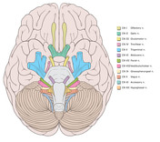
Image 3 MRI (T2 fat sat) ( update )

Image 4 MRI (T2) ( update )

Image 5 Annotated image (CN I) ( update )

Image 6 MRI (T2 fat sat) ( update )


 Unable to process the form. Check for errors and try again.