
Orbital lymphangioma
Updates to Article Attributes
Orbital lymphangiomas, also known as orbital venous lymphatic malformations, are congenital benign orbital vascular malformations composed of variable venous and lymphatic components.
Epidemiology
Orbital lymphangiomas are common in children.
Clinical presentation
- orbital swelling and proptosis: the lesion size may increase secondary to recent internal haemorrhage or upper respiratory tract infection
- orbital haemorrhage
- inflammation and cellulitis
- ocular pressure complications as optic neuropathy
Pathology
Orbital lymphangiomas are non-encapsulated venous lymphatic malformations formed of blood - or fluid - containing endothelial lacunae included within a fibrous stroma.
Radiographic features
Orbital lymphangiomas are ill-defined, poorly marginated, non-encapsulated trans-spatial multiloculated cystic and solid orbital lesions that commonly involve the whole orbital spaces including preseptal, postseptal, intraconal, and extraconal compartments.
CT
- the lesion is complex and contains cystic and solid areas. The cystic areas elicit fluid density and represent the lymphatic component of the lesion while the solid areas appear hyperdense to brain parenchyma on non-contrast CT and represent the venous component of the lesion.
- intralesional phleboliths are occasionally seen and appear as small dense foci within the solid venous part of the lesion.
- CT may demonstrate associated bony
remodelingremodelling secondary to the compressive effect of the lesion on adjacent bones
MRI
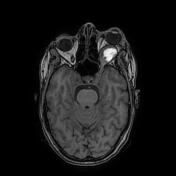
MRI is superior to CT in the evaluation of these lesions, perfectly assessing their extension and depicting various components. MRI signal is variable, depending on the fluid contents and the age of internal blood, however, it is commonly presented as follows:
- T1: iso- to hyperintense to brain parenchyma
- T2: hyperintense to brain parenchyma with occasional multiple fluid-fluid levels
- T1 C+ (Gd): marginal and septal enhancement of the cystic spaces with a variable enhancement of the solid components
Differential diagnosis
General imaging differential considerations include:
-<li>CT may demonstrate associated bony remodeling secondary to the compressive effect of the lesion on adjacent bones</li>- +<li>CT may demonstrate associated bony remodelling secondary to the compressive effect of the lesion on adjacent bones</li>
-<li><a href="/articles/orbital-cavernous-venous-malformation">cavernous hemangioma</a></li>-<li><a href="/articles/capillary-haemangioma-of-the-orbit">capillary hemangioma</a></li>- +<li><a href="/articles/orbital-cavernous-venous-malformation">cavernous haemangioma</a></li>
- +<li><a href="/articles/capillary-haemangioma-of-the-orbit">capillary haemangioma</a></li>
-<li><a href="/articles/lacrimal-gland-tumours">lacrimal gland tumors</a></li>- +<li><a href="/articles/lacrimal-gland-tumours">lacrimal gland tumours</a></li>
Image 1 Pathology (H&E) ( create )
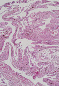
Image 2 CT (non-contrast) ( create )
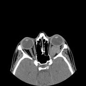
Image 3 MRI (T2) ( update )
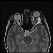
Image 4 MRI (T2 fat sat) ( update )
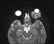
Image 5 MRI (Axial) ( update )

Image 6 MRI (T1 C+ fat sat) ( update )

Image 7 MRI (T1) ( create )


 Unable to process the form. Check for errors and try again.