
Pituicytomas are rare indolent glial tumors presumably arising from pituicytes and are only found in the neurohypophysis and infundibulum of the pituitary gland.
On this page:
Terminology
There is much confusion in the literature as to what exactly a pituicytoma is, with the term used synonymously by some authors for a number of other tumors, now thought to be separate lesions, or conversely as being distinct to other lesions, now believed to represent the same disease (e.g. pilocytic astrocytoma of the neurohypophysis, posterior pituitary astrocytoma, or infundibuloma) 1,3,4,7.
In the 5th edition (2021) of the WHO classification of CNS tumors these tumors are grouped together with granular cell tumors of the pituitary and spindle cell oncocytomas of the pituitary gland as "most likely representing a spectrum of a single nosological entity" unified by all having expression of thyroid transcription factor 1 (TTF-1) 7. Having said that, they go on to describe distinctive histological features, and as such, we continue to discuss these two other entities separately.
Epidemiology
The peak incidence of pituicytomas is between 40 and 60 years of age, although cases have been reported in a wide range of years (7-83 years of age) 6. There is a slight recognized male predilection 6.
Clinical presentation
Clinical presentation is either from endocrine dysfunction (amenorrhea, galactorrhea, infertility, diabetes insipidus, panhypopituitarism, etc.) or from compression of adjacent structures, typically the optic chiasm. Many are asymptomatic, with the lesion only found incidentally.
Pathology
Pituicytomas are classified as WHO grade 1 tumors and are usually indolent.
Macroscopic appearance
Macroscopically these tumors are well-circumscribed, soft to medium consistency with a grey to yellow homogeneous or granular cut surface. Necrosis and cystic degeneration are uncommon.
Microscopic appearance
Histologically pituicytomas are well-defined tumors composed of elongated bipolar spindle or stellate cells with rich blood supply 1,6.
Immunophenotype
Immunohistochemistry generally demonstrates the following findings 1,6:
S100: positive
vimentin: positive
TTF1: positive nuclear staining
GFAP: variable
EMA: variably/patchy
Ki-67: usually less than 2% 1,6
Radiographic features
CT
Typically pituicytomas are homogeneously enhancing, either within the pituitary fossa or in the suprasellar region. Size is variable, ranging from a few millimeters to a few centimeters.
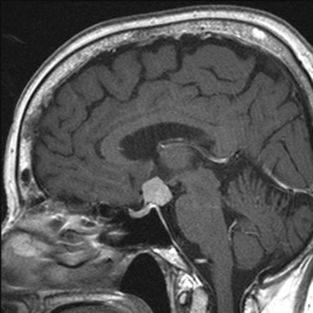
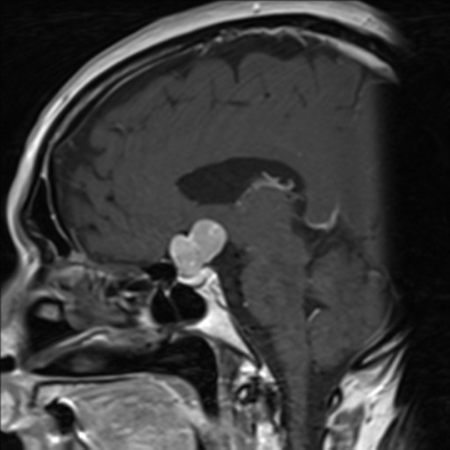
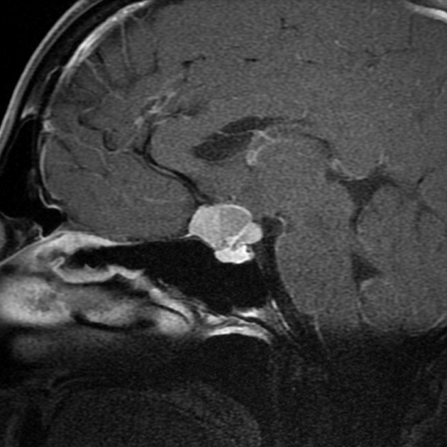
MRI
T1: isointense solid mass; posterior pituitary bright spot often absent
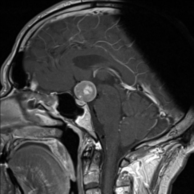
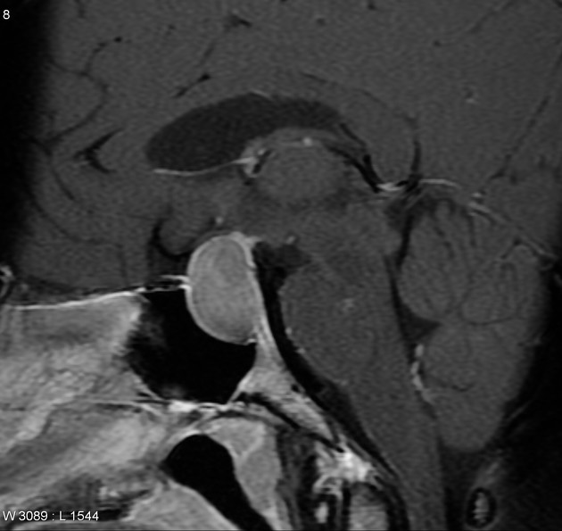
T1 C+ (Gd): bright contrast enhancement
T2: heterogeneous, hypointense to isointense
Angiography (DSA)
Pituicytomas have a rich capillary network, accounting for their usual contrast enhancement and propensity to bleed at surgery. They receive their blood supply from the normal and extensive supply to the pituitary gland, including the meningohypophyseal trunk and superior hypophyseal arteries.
Treatment and prognosis
As the tumors are benign and slow-growing if asymptomatic then expectant management is sufficient. If mass effect is present then resection may be required. Caution should be exercised as these tumors are highly vascular which may lead to difficulties with a routine transsphenoidal approach.
Complete resection can be difficult due to the structures involved and local recurrence is common 1.
Differential diagnosis
When small, and clearly localized to the infundibulum the main differential includes:
When larger, then it is difficult to anticipate the diagnosis with other diagnoses being far more common, including:
-
pituitary infiltration


 Unable to process the form. Check for errors and try again.