Thalassemia
Updates to Article Attributes
Thalassaemia is an autosomal recessive microcytic anaemia that originated in the Mediterranean region. The genetic defect causes a reduction in the rate of globin chain synthesis which causes the formation of abnormal haemoglobin molecules. The resultant anaemia is the characteristic presenting symptom of the thalassaemias.
Thalassemia is a quantitative problem of globin synthesis, whereas sickle-cell disease (a haemoglobinopathy) is a qualitative problem of synthesis of an incorrectly functioning globin.
Pathophysiology
Normal adult haemoglobin is composed of HbA (98%) and HbA2 (2%). HbA contains two α globin chains / two β globin chains, and HbA2 contains two α globin chains / two δglobin chains. They are arranged into a heterotetramer. Thalassaemia patients produce a deficiency of either α or β globin, unlike sickle-cell disease, which produces a specific mutant form of β globin.
The thalassemias are classified according to which chain of the haemoglobin molecule is affected. In α thalassemias, production of the α globin chain is reduced, while in β thalassemia production of the β globin chain is reduced.
The β globin chains are encoded by a single gene on chromosome 11; α globin chains are encoded by two closely linked genes on chromosome 16. Thus, in a normal person with two copies of each chromosome, there are two loci encoding the β chain, and four loci encoding the α chain. Deletion of one of the α loci has a high prevalence in people of African or Asian descent, making them more likely to develop α thalassemias. β thalassemias are common in Africans, but also in Greeks and Italians.
The thalassaemia trait may confer a degree of protection against malaria, which confers a selective survival advantage on carriers.
Radiographic features
Skeletal
Marrow proliferation consists of expansion of the medulla, thinning of cortical bone, and resorption of cancellous bone resulting in a generalized loss of bone density.
- skull: classic "hair-on-end" appearance
- facial bones: rodent facies
- ribs: "rib-within-a-rib" appearance, noted particularly in the middle and anterior portions of the ribs
- extramedullary
hematopoiesishaematopoiesis - premature fusion of the epiphyses
Gastrointestinal: hepatobilliary
-<li><a href="/articles/extramedullary-haematopoiesis">extramedullary hematopoiesis</a></li>- +<li><a href="/articles/extramedullary-haematopoiesis">extramedullary haematopoiesis</a></li>
-<li><a href="/articles/haemosiderosis">hemosiderosis</a></li>- +<li><a href="/articles/haemosiderosis">haemosiderosis</a></li>
References changed:
- 3. Vinay Kumar, Abul K. Abbas, Jon C. Aster. Robbins Basic Pathology. - 9. Ed. (2013) ISBN: 9781437717815 - <a href="http://books.google.com/books?vid=ISBN9781437717815">Google Books</a>
- 3. FRCPath VKMBBSMD, MBBS AKA, Aster JC. Robbins Basic Pathology. Saunders. (2012) ISBN:1437717810. <a href="http://books.google.com/books?vid=ISBN1437717810">Read it at Google Books</a> - <a href="http://www.amazon.com/gp/product/1437717810">Find it at Amazon</a><span class="auto"></span>
Image 9 CT (C+ arterial phase) ( create )
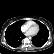
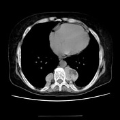
Image 10 CT (non-contrast) ( create )


 Unable to process the form. Check for errors and try again.