
Chondrosarcoma - sphenoid wing
Updates to Study Attributes
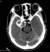
An extra-axial lesion arising from just lateral to the clivus and cavernous sinus is noted. It is of mixed density with regions of calcifications.
The location and appearance are suggestive of a chondrosarcoma. The off-midline location and ring-like calcification make a chordoma much less likely. A meningioma or trigeminal schwannoma are less likely possibilities.
Image CT (non-contrast) ( update )

Image CT (bone window) ( update )
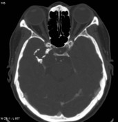
Image CT (C+ delayed) ( update )
Updates to Study Attributes
Selected MRI images confirm the presence of an extra-axial lesion arising from just lateral to the clivus and cavernous sinus. It is heterogeneous in signal with the more medial component demonstrated high T2 signal and variegated contrast enhancement, without restricted diffusion.
Features are consistent with a chondrosarcoma (histologically proven). The off-midline location and ring-like calcification make a chordoma much less likely.
Image MRI (DWI) ( update )
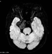
Image MRI (T1 C+) ( update )
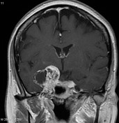
Image MRI (FLAIR) ( update )
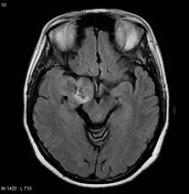
Image MRI (T1) ( update )
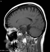
Image MRI (T1) ( update )
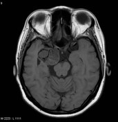
Image MRI (T1 C+) ( update )

Image MRI (T1 C+) ( update )

Image MRI (T2) ( update )
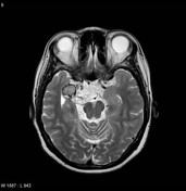
Image 1 MRI (T1) ( update )
Image 2 MRI (T1 C+) ( update )
Image 3 MRI (T2) ( update )
Image 4 MRI (FLAIR) ( update )
Image 5 MRI (T1 C+) ( update )
Image 6 MRI (T1) ( update )
Image 7 MRI (T1 C+) ( update )
Image 8 MRI (DWI) ( update )
Updates to Case Attributes
CT and MRI images demonstrate an extraaxial lesion arising from just lateralThe patient went on to the clivus and cavernous sinus. It is of mixed density on CT with regions of calcifications.
On MRI the lesion is heterogeneous in signal with the more medial component demonstrated high T2 signal and variegated contrast enhancement, without restricted diffusion.
Features are consistent withhave a chondrosarcoma (histologically proven) The off-midline location and ring-like calcification makes a chordoma much less likely.
Final Diagnosis
HIstology:Sphenoid wing
lesion, excision: Chondrosarcoma, low-grade (2/3).
Microscopic Description:
Sectionsshow a moderately cellular cartilage-forming neoplasm with large areas of tumor
tumour necrosis. The tumortumour cells are round to polygonal with hyperchromatic nuclei andare embedded in a hyaline chondromatous matrix. There are foci, however, in whichthe tumortumour cells are stellate to spindle in shape, have slightly vacuolatedcytoplasm and are embedded in a myxoid stroma. The tumortumour cells are diffusely andstrongly positive for S-100 protein and negative for EMA and cytokeratin.
Final Diagnosis: chondrosarcoma (grade 2 of 3).
-<p>CT and MRI images demonstrate an extraaxial lesion arising from just lateral to the clivus and cavernous sinus. It is of mixed density on CT with regions of calcifications. </p><p>On MRI the lesion is heterogeneous in signal with the more medial component demonstrated high T2 signal and variegated contrast enhancement, without restricted diffusion. </p><p>Features are consistent with a <a href="/articles/chondrosarcoma-of-the-base-of-skull" title="Chondrosarcoma of the base of skull">chondrosarcoma</a> (histologically proven). The off-midline location and <a href="/articles/rings-and-arcs-calcification" title="Rings and arcs calcification">ring-like calcification</a> makes a <a href="/articles/chordoma" title="Chordoma">chordoma</a> much less likely. </p><h4>Final Diagnosis:</h4> <p>Sphenoid wing-lesion, excision: Chondrosarcoma, low-grade (2/3).</p><h5>Microscopic Description: </h5><p>Sections-show a moderately cellular cartilage-forming neoplasm with large areas of tumor-necrosis. The tumor cells are round to polygonal with hyperchromatic nuclei and-are embedded in hyaline chondromatous matrix. There are foci, however, in which-the tumor cells are stellate to spindle in shape, have slightly vacuolated-cytoplasm and are embedded in myxoid stroma. The tumor cells are diffusely and-strongly positive for S-100 protein and negative for EMA and cytokeratin. </p>- +<p>The patient went on to have a resection. </p><p><strong>HIstology: </strong></p><p>Sections show a moderately cellular cartilage-forming neoplasm with large areas of tumour necrosis. The tumour cells are round to polygonal with hyperchromatic nuclei and are embedded in a hyaline chondromatous matrix. There are foci, however, in which the tumour cells are stellate to spindle in shape, have slightly vacuolated cytoplasm and are embedded in a myxoid stroma. The tumour cells are diffusely and strongly positive for S-100 protein and negative for EMA and cytokeratin.</p><p>Final Diagnosis: <a title="Chondrosarcoma" href="/articles/chondrosarcoma">chondrosarcoma</a> (<a title="Chondrosarcoma grading" href="/articles/chondrosarcoma-grading">grade 2 of 3</a>).</p>


 Unable to process the form. Check for errors and try again.