
Chondrosarcomas are a heterogeneous group of malignant cartilaginous tumors most commonly found in older patients. They can arise de novo or secondary to an existing benign cartilaginous neoplasm. On imaging, these tumors have ring-and-arc chondroid matrix mineralization with aggressive features such as lytic pattern, deep endosteal scalloping, and soft-tissue extension.
On this page:
Epidemiology
Chondrosarcomas account for 20-25% of all malignant bone tumors and are the third most common malignant bone tumor after myeloma and osteosarcoma 1,2. In patients aged >75 years, chondrosarcomas are the most common primary bone tumor 10.
Associations
Some chondrosarcomas originate from precursor lesions, namely osteochondromas or enchondromas, and are associated with 11
Diagnosis
A provisional diagnosis can often be made using a combination of clinical information, the location of the tumor, and characteristic imaging features. The diagnosis of the exact subtype and tumor grading might require histology and/or molecular pathology 1.
Clinical presentation
Patients usually present with pain, a palpable lump or a local mass effect. They are also found incidentally in imaging studies. Some chondrosarcomas present with a pathological fracture.
Hyperglycemia can occur as a paraneoplastic syndrome ref.
Pathology
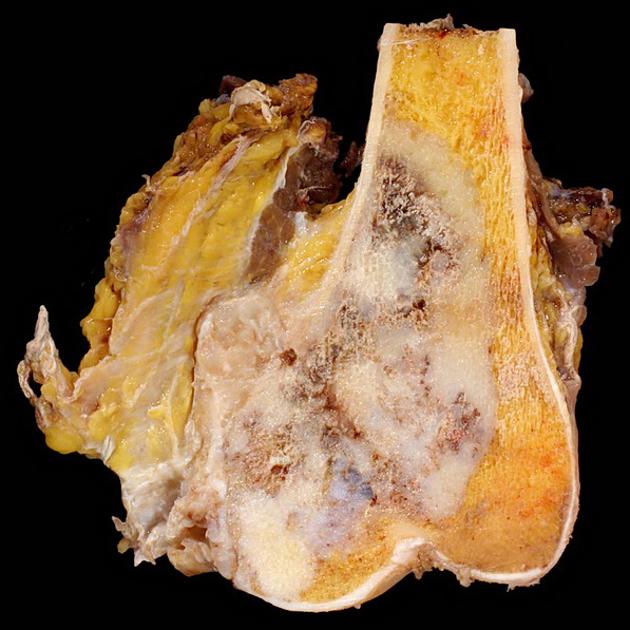
Chondrosarcomas are aggressive cartilage matrix-forming tumors and are either primary, arising de novo, or secondary originating from a pre-existent cartilaginous mass 3. Their histology can differ according to their subtype (see below).
In general, the tumors are multilobulated (due to hyaline cartilage nodules) with central high water content and peripheral endochondral ossification. On imaging, this accounts not only for the high T2 MRI signal (see below) but also for rings and arcs or popcorn calcification ref.
Subtypes
Chondrosarcomas can be classified into the following subtypes:
-
conventional chondrosarcoma (85-90%) 1,4
dedifferentiated chondrosarcoma (in up to 10% of conventional carcinomas) 3
mesenchymal chondrosarcoma (~3%) 3
periosteal chondrosarcoma (~2.5%) 3
Grading
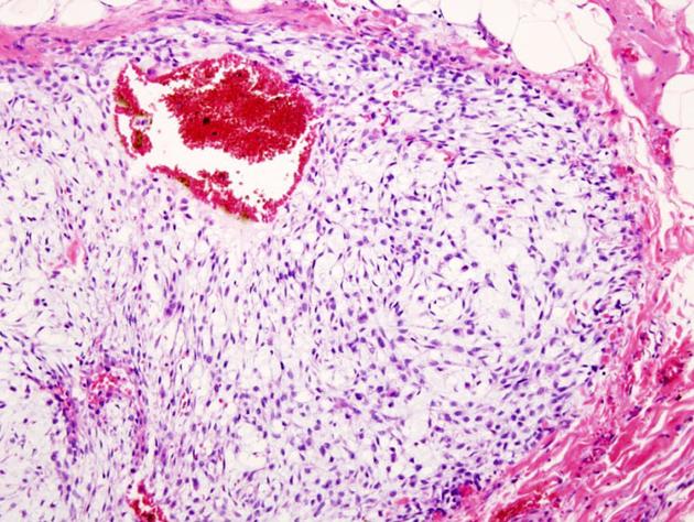
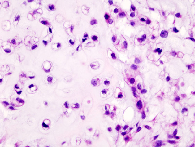
Chondrosarcomas are divided into three (sometimes four) grades based primarily on cellularity (see: chondrosarcoma grading).
Location
-
long bones: 45% (the reason is that the cartilage is more abundant in the long, tubular bones)
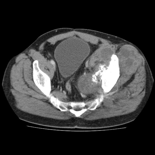
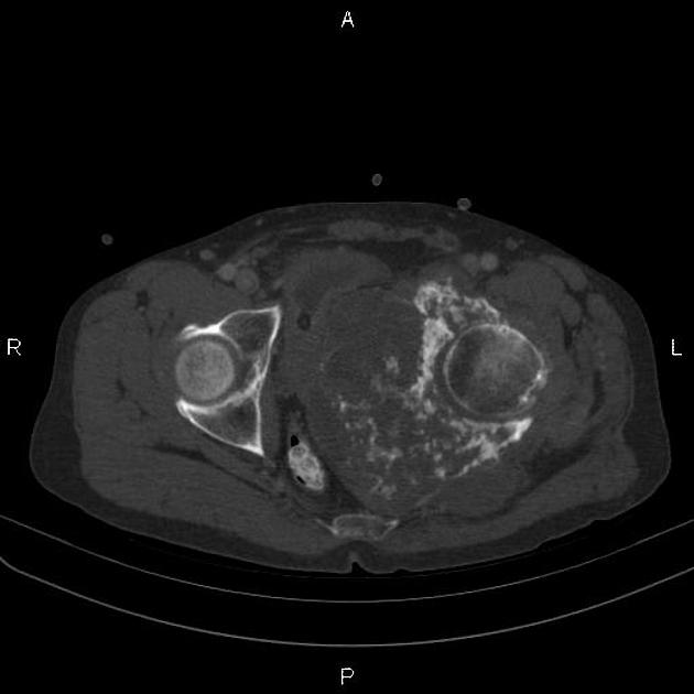
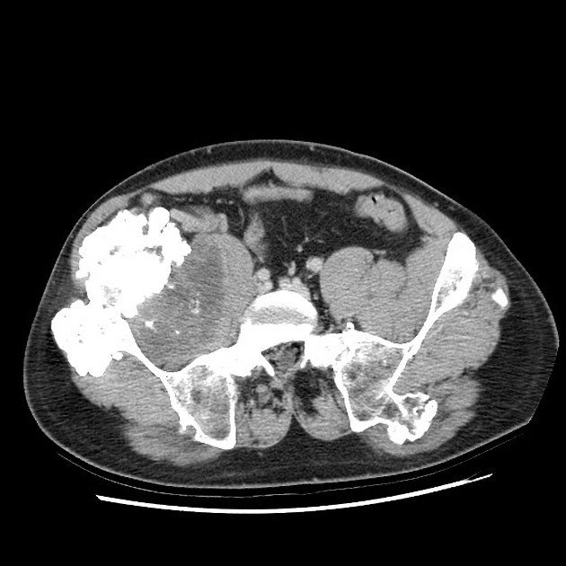
pelvis: 25% especially around the triradiate cartilage
-
ribs: 8%
patients often younger than at other sites
anterior ribs/costochondral junction
-
spine: 7%
greater male predominance 2-4:1
thoracic most common
-
location
posterior elements and vertebral body 45%
posterior elements only 40%
vertebral body only 15%
scapula: 5%
sternum: 2%
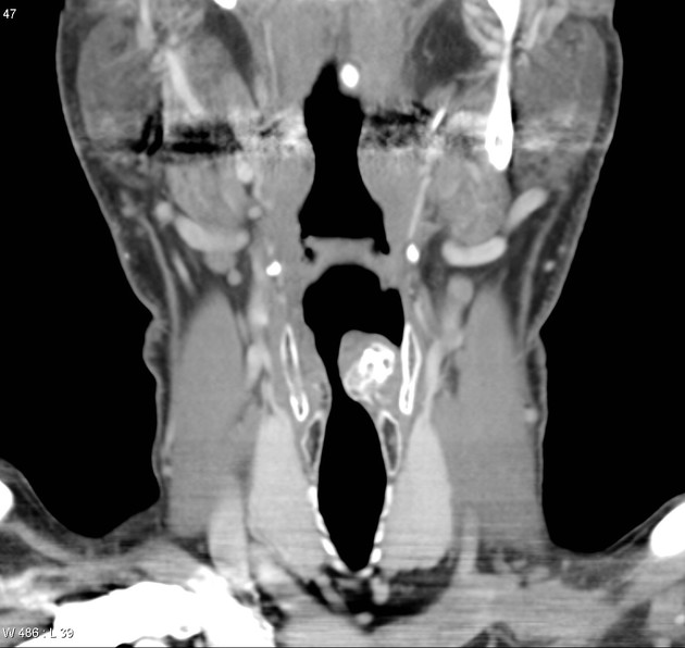
head and neck (including cervical spine): 6-7%
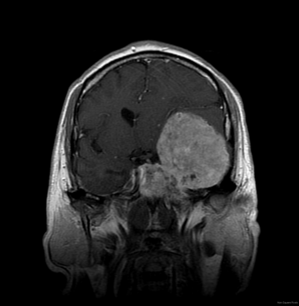
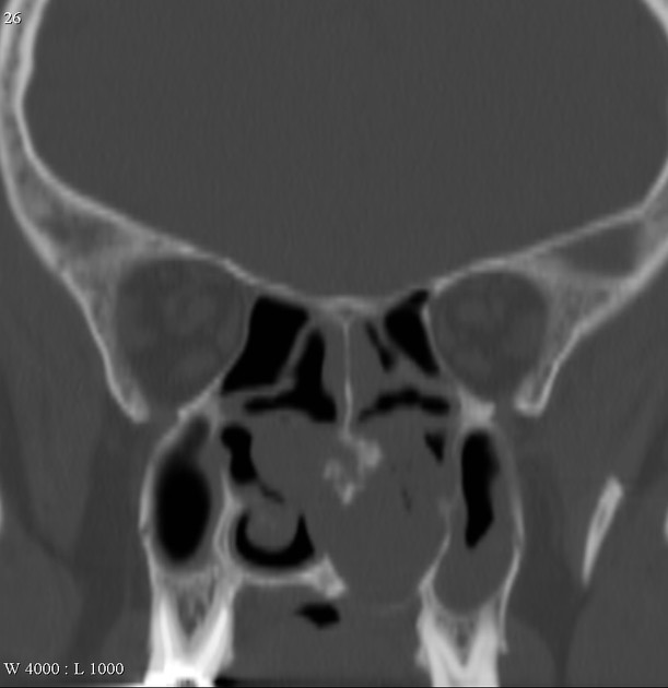
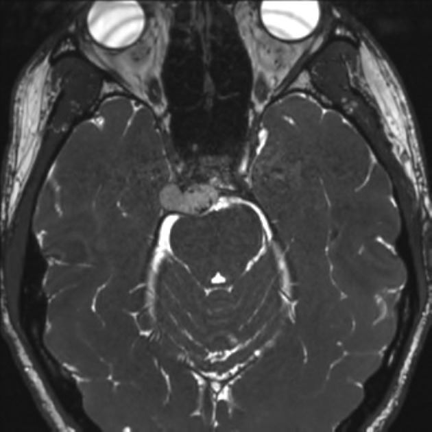
craniofacial: 2% (see chondrosarcoma of the skull base)
hands and feet: rare cf. enchondromas
Radiographic features
Imaging findings vary somewhat with different subtypes but do have some general features. Below are typical imaging appearances which are best demonstrated by conventional chondrosarcomas.
In general chondrosarcomas are large masses at the time of diagnosis, usually >4 cm in diameter and >10 cm in 50% of cases.
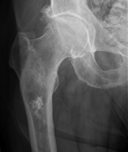
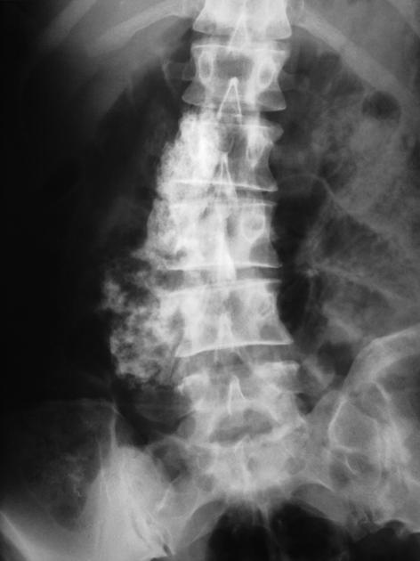
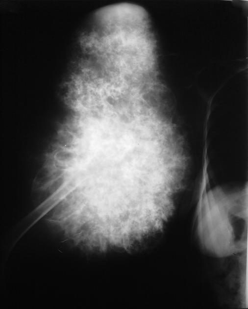
Plain radiograph
lytic (50%)
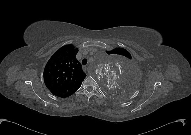
intralesional calcifications: ~70% (rings and arcs calcification or popcorn calcification)
endosteal scalloping: affecting more than two-thirds of the cortical thickness (cf. less than two-thirds in enchondromas)
moth-eaten appearance or permeative appearance in higher grade tumors (see chondrosarcoma grading), e.g. myxoid, dedifferentiated and mesenchymal chondrosarcomas
cortical remodeling, thickening and periosteal reaction are also useful in distinguishing between an enchondroma and low-grade chondrosarcoma (see enchondroma vs low-grade chondrosarcoma)
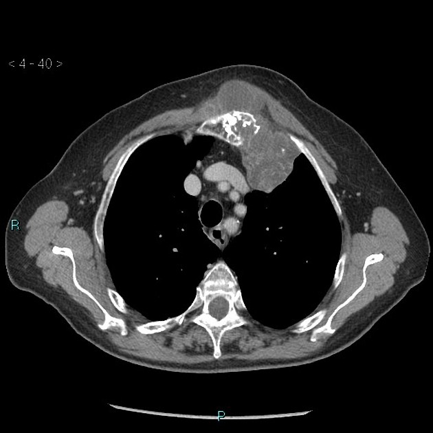
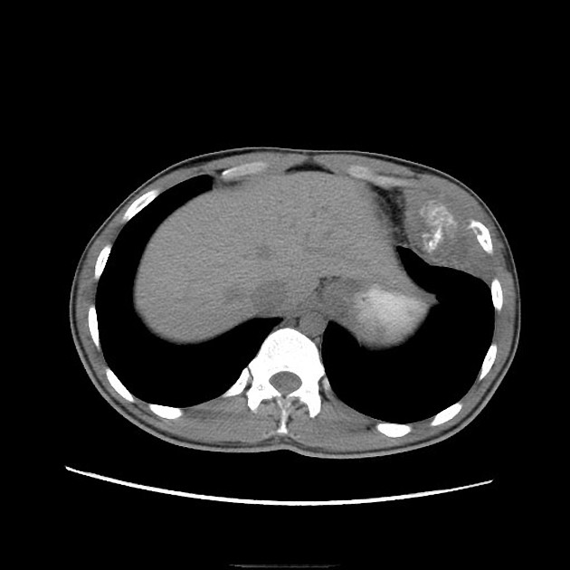
CT
The features seen on CT are the same as on plain film but are simply better seen:
~95% of cases demonstrate matrix calcification, cf. ~70% (range 60-78% on x-ray)
cortical breach, seen in ~90% of long bone chondrosarcoma, cf. only ~10% of enchondromas
soft tissue mass: tumor cellularity, and therefore density, increases with the increased grade of the tumor
heterogenous contrast enhancement
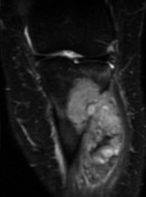
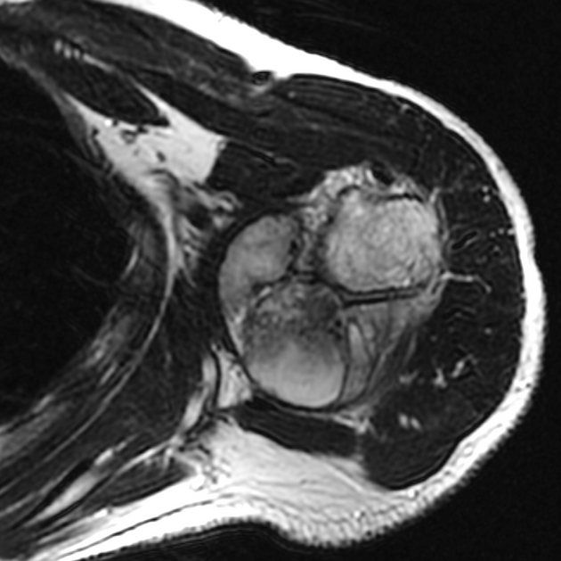
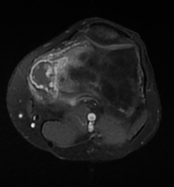
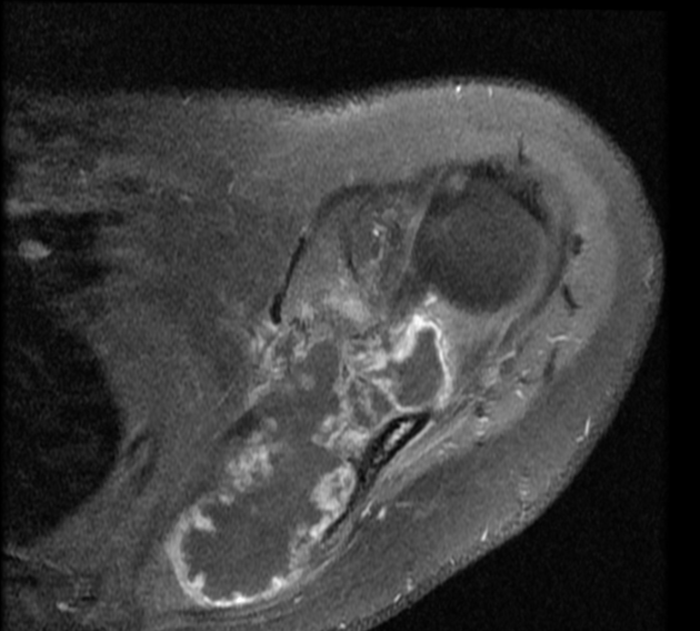
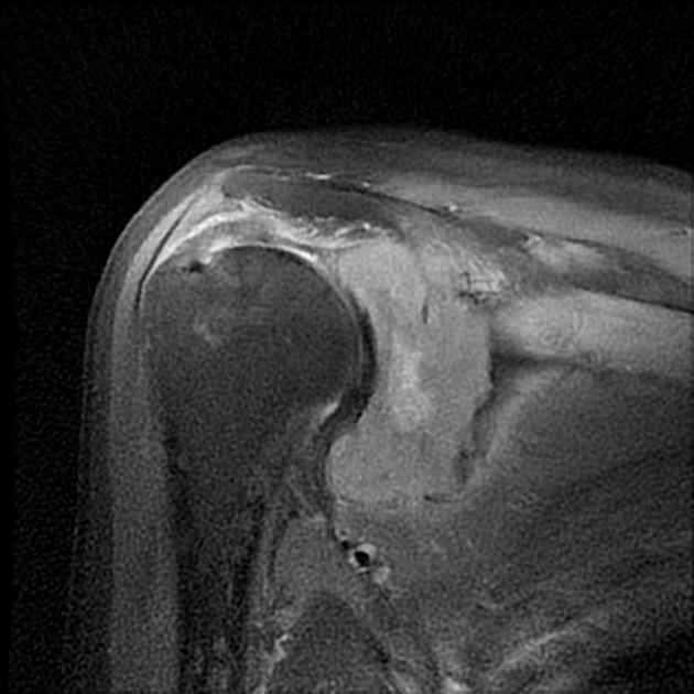
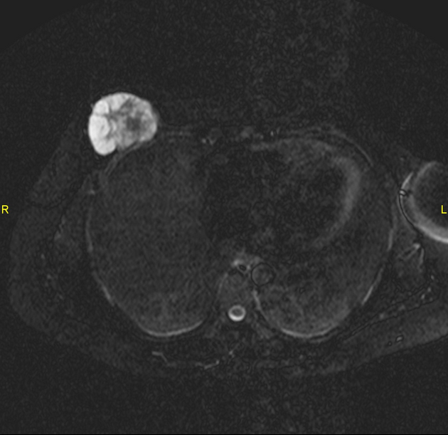
MRI
-
T1: low to intermediate signal
iso- to slightly hyperintense cf. muscle
iso- to slightly hypointense cf. grey matter (see chondrosarcoma of the base of the skull)
T2: very high intensity in non-mineralized/calcified portions - the cartilage is a hydrophilic tissue with high water content 6
gradient echo/SWI: blooming of mineralized/calcified portions
-
T1 C+ (Gd)
enhancement can be septal and peripheral rim-like corresponding to fibrovascular septation between lobules of hyaline cartilage - ring-and-arc enhancement pattern 6,7
most demonstrate heterogeneous moderate to intense contrast enhancement ref
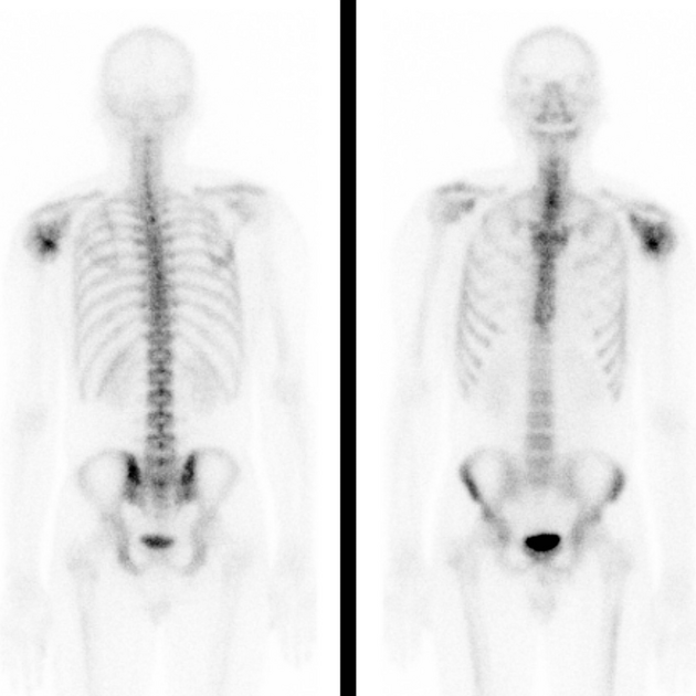
Nuclear medicine
Bone scintigraphy
Typically chondrosarcomas demonstrate increased uptake on bone scintigraphy, seen in over 80% of cases, and usually, the uptake is quite intense ref. This is helpful to distinguish low-grade chondrosarcoma from an enchondroma as the latter has increased uptake in ~20% of cases, and usually to a lesser degree (see: enchondroma vs low-grade chondrosarcoma) ref.
Treatment and prognosis
The management and prognosis depend on the subtype, the histological grade and the location of the tumors (see also respective articles). Treatment usually involves surgery, and the type of excision will again vary with the type, grade and location of the tumor 2. Chemotherapy and radiation generally play no role, except for mesenchymal chondrosarcomas 2,3.


 Unable to process the form. Check for errors and try again.