
Cases
By sharing our collective experience through interesting and classic patient cases, we can make a real difference in how people are imaged and diagnosed. Each case belongs to a contributing member and all cases are reviewed by our dedicated editors to ensure they reach quality standards and abide by privacy guidelines. Cases can public or unlisted and then be viewed directly or added to articles, playlists or multiple choice questions. Find out more about cases.


58,983 results found
Case
CTEV with Arnold-Chiari malformation

Published
11 Jun 2024
79% complete
Ultrasound
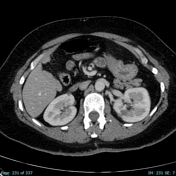
Case
Renal replacement lipomatosis

Published
11 Jun 2024
77% complete
CT
Case
Acromial type II - curved
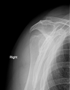
Published
11 Jun 2024
88% complete
X-ray
Case
Cervical esophageal carcinoma (neck ultrasound)

Published
11 Jun 2024
79% complete
Ultrasound
Case
Papillary process of the caudate lobe in cirrhotic patient
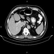
Published
11 Jun 2024
92% complete
CT
Case
Humeral surgical neck fracture
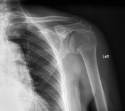
Published
11 Jun 2024
88% complete
X-ray
Case
Primary intraventricular hemorrhage
Published
11 Jun 2024
59% complete
CT
Case
Double J stent perforating renal parenchyma

Published
11 Jun 2024
95% complete
CT
Case
Parafalcine empyema

Published
11 Jun 2024
95% complete
CT
Case
Splenic artery pseudoaneurysm mimicking pancreatic mass

Published
11 Jun 2024
92% complete
DSA (angiography)
MRI
CT
Case
Mineralizing angiopathy involving lenticulostriate arteries

Published
11 Jun 2024
89% complete
CT
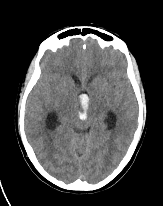
Case
Hypoxic-ischemic brain injury
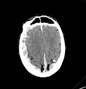
Published
11 Jun 2024
95% complete
CT
Case
Hepatic hydatidosis

Published
11 Jun 2024
74% complete
CT
Case
Nasolacrimal duct obstruction

Published
11 Jun 2024
95% complete
CT
Case
Normal bilateral foot weight-bearing DP view

Published
11 Jun 2024
75% complete
X-ray
Case
Hiatus hernia

Published
10 Jun 2024
72% complete
X-ray
Case
Intestinal nonrotation
Published
10 Jun 2024
89% complete
CT
Case
Pigmented villonodular synovitis (PVNS)
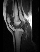
Published
10 Jun 2024
68% complete
MRI
Case
Complication of gastric banding resulting in acute small bowel obstruction

Published
10 Jun 2024
92% complete
CT
Case
Myocardial bridging of the LAD

Published
10 Jun 2024
92% complete
CT
ADVERTISEMENT: Supporters see fewer/no ads


 Unable to process the form. Check for errors and try again.