


5,898 results found
Case
3 T v 1.5 T MRI prostate with total hip prosthesis

Published
16 Dec 2017
92% complete
MRI
Case
AAST grade IV kidney injury with CEUS follow-up

Published
21 Nov 2019
98% complete
CT
Ultrasound
Article
AAST kidney injury scale
The American Association for the Surgery of Trauma (AAST) renal injury scale, updated in 2018, is the most widely used grading system for renal trauma.
The 2018 update incorporates "vascular injury" (i.e. pseudoaneurysm, arteriovenous fistula) into the imaging criteria for visceral injury.
Cla...
Article
Abdomen (KUB view)
The kidneys, ureters, bladder (KUB) radiograph is optimized for assessment of the urogenital system, and should not be confused with the AP supine abdomen view. However, in cases where the patient may have both gastrointestinal and urogenital abnormalities, all pathologies will still be reported...
Article
Abdomen (oblique view)
AP oblique supine radiograph is a projection often used in barium studies and foreign body localization.
Indications
This view is normally performed when localizing foreign bodies or lines within the abdominal cavity. Additionally, the oblique abdominal series can be utilized in the assessment...
Article
Abdomen (PA prone view)
The PA prone radiograph is rarely performed and is often utilized when a patient is unable to lay supine. The projection is adequate for the examination of the abdominal cavity, however, not as practical for the renal structures due to magnification.
Indications
This view is useful in visualiz...
Article
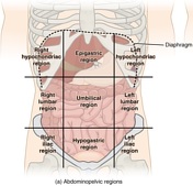
Abdominal and pelvic anatomy
Abdominal and pelvic anatomy encompasses the anatomy of all structures of the abdominal and pelvic cavities.
This anatomy section promotes the use of the Terminologia Anatomica, the international standard of anatomical nomenclature.
Case
Abdominal aorta and retroperitoneum (Gray's illustration)

Published
17 Jul 2017
38% complete
Diagram
Case
Abdominal calcifications

Published
19 Jul 2016
89% complete
CT
X-ray
Annotated image
Case
Abdominal multi-trauma - devascularised kidney and liver, spleen and pancreatic lacerations

Published
28 Aug 2015
80% complete
CT
Case
Abdominal multitrauma - pediatric

Published
13 Aug 2020
95% complete
CT
Annotated image
Article
Abdominal radiography
Abdominal radiography can be useful in many settings. Before the advent of CT, it was a primary means of investigating gastrointestinal pathology and often allowed indirect evaluation of other abdominal viscera.
Indications
Although abdominal radiography has lower sensitivity and specificity t...
Case
Abdominal surface anatomy (creative commons illustration)
Published
20 Mar 2018
35% complete
Diagram
Article
Abdominal tuberculosis
Abdominal tuberculosis can manifest in almost every abdominopelvic organ:
gastrointestinal tuberculosis
esophageal tuberculosis
gastric tuberculosis
duodenal tuberculosis
jejunal and ileal tuberculosis
ileocecal tuberculosis
colorectal tuberculosis
tuberculous pe...
Case
Abdominopelvic hydatid cyst

Published
28 May 2022
77% complete
CT
Case
Aberrant extra renal artery and vein arising from common iliac vessels

Published
22 Feb 2024
86% complete
CT
Case
Aberrant renal artery

Published
14 Aug 2014
75% complete
Ultrasound
Article
Abnormal renal rotation
Abnormal renal rotation, also known as renal malrotation, refers to an anatomical variation in the position of the kidneys, in particular to anomalous orientation of the renal hilum. It may occur unilaterally or bilaterally. It is almost always an asymptomatic incidental finding.
Epidemiology
...
Case
Abnormal renal vascular impression on intravenous pyelogram

Published
07 May 2012
60% complete
X-ray
Article
Abnormal testicular Doppler flow (differential)
Abnormal testicular Doppler flow (arterial, venous, or both) can be a differential challenge. Always remember that the patient's presenting history helps quite a bit in narrowing the differential.
Reduced flow
partial testicular torsion (<360 degrees)
venous outflow is obstructed first, resul...


 Unable to process the form. Check for errors and try again.