
Items tagged “knee”
419 results found
Case
Lipoma arborescens

Published
05 Jan 2014
48% complete
MRI
Article
Knee menisci
The knee menisci are fibrocartilaginous structures that sit within the knee joint, deepening the tibiofemoral articulation. Their main role is shock absorption, improve stability of the knee joint, and load transmission. They also play an important role in synovial fluid dynamic circulation and ...
Case
Posterior knee anatomy: Gray's anatomy illustration

Published
09 Jan 2014
29% complete
Diagram
Case
Osteoarthritis of the knee

Published
18 Jan 2014
94% complete
X-ray
Article
Lateral patellar dislocation
Lateral patellar dislocation refers to lateral displacement followed by dislocation of the patella due to disruptive changes to the medial patellar retinaculum.
Epidemiology
Patellar dislocation accounts for ~3% of all knee injuries and is commonly seen in those individuals who participate in ...
Case
Physeal anatomy - illustration
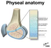
Published
31 Jan 2014
29% complete
Diagram
Case
Tibial spine fracture with anterior cruciate ligament avulsion - type 4

Published
31 Jan 2014
95% complete
X-ray
CT
Article
Intercondylar area
The intercondylar area is the rough, central part of the tibial plateau.
Gross anatomy
The intercondylar area is located between the proximal articular surfaces of the medial and lateral tibial condyles. It is non-articular. In the middle of the intercondylar area are:
intercondylar eminenc...
Article
Tibial plateau
The tibial plateau (plural: plateaus or plateaux are equally acceptable 4) is the proximal articular surface of the tibia.
Terminology
Strictly the plateau refers to the whole articular surface of the proximal tibia. Therefore, saying "medial tibial plateau" or "lateral tibial plateau", or, ev...
Case
Medial patellar plica syndrome

Published
02 May 2014
74% complete
MRI
Case
Bucket handle tear - medial meniscus

Published
13 May 2014
68% complete
MRI
Case
Secondary synovial osteochondromatosis and enchondroma

Published
21 Jun 2014
79% complete
X-ray
Case
Remote ganglion cyst and Hoffa impingement

Published
27 Jul 2014
77% complete
MRI
Diagram
Article
Wiberg classification of patella shape
Wiberg classification is a system used to describe the shape of the patella based mainly on the asymmetry between the patellar medial and lateral facets on axial views of the patella. Increasing number type indicates a larger degree of asymmetry.
Classification
type I (or type A)
roughly symm...
Case
Meniscal ossicle

Published
08 Aug 2014
92% complete
X-ray
MRI
Case
Rice bodies - knee synovial chondromatosis

Published
20 Sep 2022
90% complete
X-ray
MRI
Article
Knee joint
The knee joint is a modified hinge joint between the femur, tibia, and patella. It is the largest synovial joint in the body and allows flexion and extension of the leg as well as some rotation in the flexed position.
Summary
location: two condylar joints between femur and tibia; saddle joint ...
Article
Medial collateral ligament of the knee
The medial collateral ligament (MCL) of the knee is a flat, triangular band on its medial aspect that resists valgus forces. It forms part of the medial capsuloligamentous complex of the knee.
Gross anatomy
The medial collateral ligament measures 8-10 cm in length and has superficial and deep...
Article
Lateral collateral ligament of the knee
The lateral (fibular) collateral ligament (LCL) is on the lateral aspect of the knee and forms part of the posterolateral corner. It is a major knee stabilizer against varus forces 6. The lateral aspect of the knee is divided into three layers and the LCL is part of the deep layer of the latera...
Case
Bipartite patella

Published
01 Nov 2014
84% complete
X-ray
CT


 Unable to process the form. Check for errors and try again.