


305 results found
Case
Coracoacromial ligament ossification

Published
13 Feb 2013
65% complete
X-ray
Case
Pes anserinus spur

Published
02 Feb 2013
47% complete
X-ray
Case
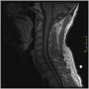
Vertebral hemangioma involving dens
Published
31 Jan 2013
59% complete
MRI
Case
Osteochondritis dissecans of the talus

Published
31 Jan 2013
95% complete
MRI
Case
Diagram - intracranial hemorrhage

Published
30 Jan 2013
44% complete
Diagram
Case
Normal marrow conversion (diagram)

Published
30 Jan 2013
35% complete
Diagram
Case
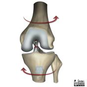
Anterior cruciate ligament injury (illustrations)
Published
23 Jan 2013
41% complete
Diagram
Case
Liposclerosing myxofibrous tumor

Published
16 Jan 2013
52% complete
X-ray
Annotated image
Case
Medial tibial stress syndrome

Published
16 Jan 2013
82% complete
X-ray
Annotated image
Case
Osteopoikilosis (MRI)

Published
15 Jan 2013
92% complete
MRI
Case
Fredericson MRI classification of medial tibial stress syndrome

Published
14 Jan 2013
41% complete
Diagram
Case
Pelvic insufficiency fractures (illustrations)

Published
28 Dec 2012
38% complete
Diagram
Case
Bosniak classification of renal cysts (illustrations)

Published
26 Dec 2012
41% complete
Diagram
Case
Diagram - Bismuth-Corlette classification of perihilar cholangiocarcinoma

Published
22 Dec 2012
41% complete
Diagram
Case
Diagram - Salter-Harris fracture: type 2

Published
19 Dec 2012
29% complete
Diagram
Case
Diagram - distribution of solitary enchondromas of the hand

Published
19 Dec 2012
41% complete
Diagram
Case
Skier's thumb - type I

Published
01 Nov 2012
63% complete
Diagram
Case
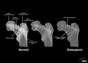
Illustration of Ward's triangle
Published
25 Oct 2012
29% complete
Diagram
Case
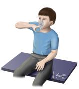
Illustration of scurvy signs

Published
25 Oct 2012
16% complete
Diagram
Case
Radioulnar synostosis - illustration of functional difficulties
Published
24 Oct 2012
29% complete
Diagram
Case
Chondrocalcinosis: diagram of causes

Published
01 Oct 2012
32% complete
Diagram
Case
Accessory ossicle of the anterior arch of the atlas

Published
27 Sep 2012
74% complete
Diagram
X-ray
Case
Illustration of a "dural tail" sign

Published
03 Aug 2012
32% complete
Diagram
ADVERTISEMENT: Supporters see fewer/no ads


 Unable to process the form. Check for errors and try again.